Comparative Study
. 2005 Jul 12;102(28):9766-71.
doi: 10.1073/pnas.0503891102. Epub 2005 Jun 28.

Sequential phosphorylation of CCAAT enhancer-binding protein beta by MAPK and glycogen synthase kinase 3beta is required for adipogenesis

Affiliations
Comparative Study

Sequential phosphorylation of CCAAT enhancer-binding protein beta by MAPK and glycogen synthase kinase 3beta is required for adipogenesis

Qi-Qun Tang et al. Proc Natl Acad Sci U S A. .

Abstract

CCAAT enhancer-binding protein (C/EBP)beta, C/EBPalpha, and peroxisome proliferator activated receptor (PPAR)gamma act in a cascade where C/EBPbeta activates expression of C/EBPalpha and PPARgamma, which then function as pleiotropic activators of genes that produce the adipocyte phenotype. When growth-arrested 3T3-L1 preadipocytes are induced to differentiate, C/EBPbeta is rapidly expressed but still lacks DNA-binding activity. After a long (14-hour) lag, glycogen synthase kinase 3beta enters the nucleus, which correlates with hyperphosphorylation of C/EBPbeta and acquisition of DNA-binding activity. Concurrently, 3T3-L1 preadipocytes synchronously enter S phase and undergo mitotic clonal expansion, a prerequisite for terminal differentiation. Ex vivo and in vitro experiments with C/EBPbeta show that phosphorylation of Thr-188 by mitogen-activating protein kinase "primes" C/EBPbeta for subsequent phosphorylation on Ser-184 and Thr-179 by glycogen synthase kinase 3beta, acquisition of DNA-binding function, and transactivation of the C/EBPalpha and PPARgamma genes. The delayed transactivation of the C/EBPalpha and PPARgamma genes by C/EBPbeta appears necessary to allow mitotic clonal expansion, which would otherwise be prevented, because C/EBPalpha and PPARgamma are antimitotic.


Figures

Fig. 1.
Fig. 1.
Changes in phosphorylation state and DNA-binding activity of C/EBPβ during differentiation. Two-day postconfluent 3T3-L1 preadipocytes were induced to differentiate nuclear extracts prepared at 4, 16, and 24 h after induction (treated or not with alkaline phosphatase) and then subjected to (A) 2D gel analysis and immunoblotting with anti-C-terminal C/EBPβ antibody and (B) EMSA to assess DNA-binding activity using a oligonucleotide probe corresponding to the C/EBP regulatory element in the proximal promoter of the C/EBPα gene.
Fig. 2.
Fig. 2.
Expression and phosphorylation of C/EBPβ and MAPK during differentiation. (A–C) Two-day postconfluent 3T3-L1 preadipocytes were induced to differentiate. At the times indicated, cell extracts were prepared and immunoblotted with antibodies directed against (A) the C-terminal peptide of C/EBPβ, (B) phospho-Thr-188 C/EBPβ, and (C) phospho-MAPK (Upper) and MAPK (Lower). (D) Transactivation of a C/EBPα promoter-luciferase reporter gene by WT or mutant (Thr-188→ Ala) C/EBPβ expression vector.
Fig. 3.
Fig. 3.
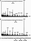
Identification of phosphorylation sites in C/EBPβ during differentiation. (A and B) Nuclear extracts were prepared before and 24 h after induction of differentiation. C/EBPβ was isolated and separated by SDS/PAGE. The band containing C/EBPβ (38 kDa) was excised from the gel, digested by trypsin, and analyzed by automated nanoLC-MS/MS as described (26). Ions labeled with an asterisk were generated from the peptide in which the phosphoserine and phosphothreonine were converted into dehydroalanine and dehydroaminobutyric acid by β-elimination, respectively. (A) Partial MS/MS spectrum of one of the species in which the phosphorylation site was localized to Thr-179. (B) Partial MS/MS spectrum of the second species where the phosphorylation site was localized to Ser-184. (C) Amino acid sequence of the tryptic peptide identifying the phosphorylation sites in Fig. 2B (Thr-188) and in A (Thr-179) and B (Ser-184) above.
Fig. 4.
Fig. 4.
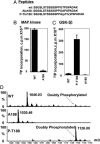
Phosphorylation of WT and mutant peptides by MAPK and GSK3β. (A) Amino acid sequences of the WT and mutant (Thr-188→ Ala and Thr-188→phospho-Thr-188) synthetic peptides used as substrates for MAPK (B) and GSK3β (C) below. Peptides were incubated individually with MAPK or GSK3β and [γ-32P]ATP/Mg, after which 32P-incorporation into the peptide was determined. (D) MS analysis of WT and mutant peptides phosphorylated in vitro by GSK3β. WT, WT peptide; Ala-188, Thr-188→ Ala peptide; and P-Thr-188, phospho-Thr-188 peptide.
Fig. 5.
Fig. 5.
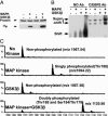
In vitro phosphorylation of full-length C/EBPβ by MAPK and GSK3β leads to acquisition of DNA-binding activity. (A)[32P] labeling of WT C/EBPβ, (B) DNA-binding activity assessed by EMSA, and (C) MS analysis of C/EBPβ after in vitro phosphorylation by MAPK and/or GSK3β. After incubation with MAPK and/or GSK3β and ATP, recombinant full-length GST-C/EBPβ(LAP) was digested by trypsin and analyzed by MS. The masses (m/z) of tryptic peptide products are shown.
Fig. 6.
Fig. 6.
Mutation of Thr-188, Ser-184, and Thr-179 to Ala abolishes DNA-binding and transactivation activities of C/EBPβ. (A) recombinant WT or AAA mutant C/EBPβ was incubated with MAPK and GSK3β after which [32P] labeling and DNA-binding activity (by EMSA) of C/EBPβ were assessed. (B) Transactivation of a C/EBPα promoter-luciferase reporter gene by WT or AAA mutant (Thr-188, Ser-184, Thr-179→ Ala) C/EBPβ expression vector.
Fig. 7.
Fig. 7.
Inhibition of MAPK or GSK3β disrupts DNA-binding activity of C/EBPβ and differentiation. U0126 (20 μM, MAPK inhibitor) or SB216763 (20 μM, GSK3β inhibitor) was added 60 min before and at the time of induction. (A) Twenty-four hours after induction, DNA-binding activity of C/EBPβ was determined by EMSA, and (B) ChIP analysis was performed with oligonucleotide primers bracketing the C/EBP-binding site in the 422/aP2 gene promoter. (C) On day 8, cells were stained with Oil-red O, and (D) expression of C/EBPβ was assessed by Western blotting on day 1 and adipocyte markers (C/EBPα, PPARγ, and 422/aP2) on day 6.

References

    1. Tang, Q.-Q., Otto, T. C. & Lane, M. D. (2003) Proc. Natl. Acad. Sci. USA 100, 850–855. - PMC - PubMed
    1. Zhang, J. W., Tang, Q.-Q., Vinson, C. & Lane, M. D. (2004) Proc. Natl. Acad. Sci. USA 101, 43–47. - PMC - PubMed
    1. Christy, R. J., Kaestner, K. H., Geiman, D. E. & Lane, M. D. (1991) Proc. Natl. Acad. Sci. USA 88, 2593–2597. - PMC - PubMed
    1. Tang, Q.-Q., Jiang, M.-S. & Lane, M. D. (1999) Mol. Cell. Biol. 19, 4855–4865. - PMC - PubMed
    1. Zhu, Y., Qi, C., Korenberg, J. R., Chen, X. N., Noya, D., Rao, M. S. & Reddy, J. K. (1995) Proc. Natl. Acad. Sci. USA 92, 7921–7925. - PMC - PubMed

Publication types

MeSH terms

Substances

LinkOut - more resources