. 2005 Jul 15;19(14):1674-9.
doi: 10.1101/gad.1334005. Epub 2005 Jun 28.

Dicer-1 and R3D1-L catalyze microRNA maturation in Drosophila

Affiliations

Dicer-1 and R3D1-L catalyze microRNA maturation in Drosophila

Feng Jiang et al. Genes Dev. .

Abstract

In Drosophila melanogaster, Dicer-2/R2D2 and Dicer-1 generate small interfering RNA (siRNA) and microRNA (miRNA), respectively. Here we identify a novel dsRNA-binding protein, which we named R3D1-L, that forms a stable complex with Dicer-1 in vitro and in vivo. While depletion of R3D1-L by RNAi causes accumulation of precursor miRNA (pre-miRNA) in S2 cells, recombinant R3D1-L enhances miRNA production by Dicer-1 in vitro. Furthermore, R3D1 deficiency causes miRNA-generating defect and severe sterility in male and female flies. Therefore, R3D1-L functions in concert with Dicer-1 in miRNA biogenesis and is required for reproductive development in Drosophila.


Figures
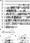
Figure 1.
Figure 1.
(A, top) A sequence alignment of R3D1-L, R2D2, and RDE-4. Boxes refer to the most conserved motifs of three putative dsRNA-binding domains. (Bottom) The underlined region represents 46 amino acids encoded by exon 4 that is alternatively spliced to create R3D1-L and R3D1-S. (B) A schematic diagram showing alternative splicing of R3D1-L and R3D1-S. Exons are shown as boxes with numbers inside. (C) Immunoprecipitations (IPs) were performed in S100 of S2 cells with anti-Dicer-1 or anti-Dicer-2 antibodies followed by Western blot using anti-R3D1 or anti-AGO1 antibodies. (D) IPs were performed with anti-R2D2 or anti-R3D1 antibodies followed by Western blots with corresponding antibodies to detect Dicer-1, Dicer-2, and AGO1. S100 lanes represent 5% input for IPs.
Figure 2.
Figure 2.
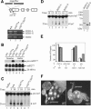
(AC) Purification of the miRNA-generating activity from S2 cell extracts by a five-step chromatography procedure (Supplemental Material). (Top) One microliter of each fraction was used for the pre-miRNA-processing assay. (Bottom) Western blots were performed with corresponding antibodies to detect Dicer-1, R3D1, Dicer-2, and R2D2 following Mono S (A), Mono Q (B), and Hydroxyapatite (C) columns. The fractions 21–23 from the Mono S column were pooled and loaded onto the Mono Q column, whereas the fractions 23–24 from the Mono Q column were pooled and loaded onto the Hydroxyapatite column. The asterisks in Dicer-1 blots represent a cross-reacting protein or a degradation product of Dicer-1. Neither Dicer-2 nor R2D2 was detected in C (data not shown).
Figure 3.
Figure 3.
(A) Western blots were performed with corresponding antibodies to measure the levels of GFP, AGO1, Dicer-1, Dicer-2, and R3D1 in S2/GFP cells following various RNAi treatments (see information of various dsRNA in Supplementary Fig. 1). The asterisk refers to R3D1-S and a comigrating cross-reacting protein. (B) Northern blots were performed to measure the levels of pre-bantam and bantam miRNA. 2S rRNA was used as loading control.
Figure 4.
Figure 4.
(A) A Coomassie-stained SDS–polyacrylamide gel (PAGE) showing purified recombinant Dicer-1, R3D1-L, and Dicer-1/R3D1-L complex. (B) Dicer-1 and Dicer-2 display different substrate specificities. The pre-miRNA-processing (top) and siRNA-generating (bottom) assays were performed with various amounts of Dicer-1 (lanes 25) or Dicer-2 (lanes 69) (Liu et al. 2003). (C) Dicer-1 and Dicer-2 have different ATP requirements. The pre-miRNA-processing assays were performed with various amounts of Dicer-1 (top) or Dicer-1/R3D1-L (bottom) in the absence (lanes 25) or presence (lanes 69) of ATP. (D) R3D1-L enhances miRNA production by Dicer-1. The pre-miRNA-processing assays were performed with 25 pmol Dicer-1 alone (lane 2) or in combination with 30, 100, or 300 pmol R3D1-L (lanes 35), or 300 pmol R3D1-L (lane 6). Lane 1 is a 21-nt marker. (E) Mg2+ is required for Dicer-1 cleavage of pre-miRNA. The pre-miRNA-processing assays were performed with buffer alone (lane 1), or with 1 nmol Dicer-1 in 10 mM Mg2+ (lane 2) or 0.2 mM Mg2+ and 2.5 mM EDTA (lane 3). (F) R3D1-L increases Dicer-1's affinity for pre-miRNA. The gel-shift assays were performed without Mg2+ in buffer alone (lane 1) or with 1 nmol Dicer-1 alone (lane 2) or in combination with 1, 3, or 10 nmol R3D1-L (lanes 35), and 10 nmol R3D1-L (lane 6).
Figure 5.
Figure 5.
(A, top) A schematic diagram of the insertion site of piggyBac. (Bottom) The mRNA levels of R3D1-L, R3D1-S, and R2D2 were measured by semiquantitative RT–PCR in wild-type, heterozygous, and homozygous r3d1PB female flies. (B) The levels of pre-miR277 and miR277 were measured by Northern blots in wild-type, heterozygous, and homozygous r3d1PB female (lanes 13) and male (lanes 46) flies. (C) The pre-miRNA-processing assays were performed with 5 μg whole fly extracts prepared from wild-type (lanes 2,3), heterozygous (lanes 4,5), and homozygous (lanes 6,7) r3d1PB female flies. (D, left) The pre-miRNA-processing assays were performed with 10 μg r3d1PB/r3d1PB mutant extracts alone (lanes 1,2) or in combination with 35 ng of recombinant R3D1-L (lanes 3,4) or R3D1-S (lanes 5,6). (Right) A Coomassie-stained SDS-PAGE showing purified recombinant R3D1-L and R3D1-S. (E) A graph showing infertility of homozygous r3d1PB male and female flies. Three mating vials were set up for each cross (shown below the X-axis). The numbers of progeny are shown on the Y-axis. (F) Images of a wild-type (left) or r3d1PB/r3d1PB mutant (right) ovariole stained with Hoechst dye to reveal the nuclei. (tf) Terminal filament.

References

    1. Aravin A.A., Lagos-Quintana, M., Yalcin A., Zavolan, M., Marks, D., Snyder, B., Gaasterland, T., Meyer, J., and Tuschl, T. 2003. The small RNA profile during Drosophila melanogaster development. Dev. Cell 5: 337–350. - PubMed
    1. Bartel D.P. 2004. MicroRNAs: Genomics, biogenesis, mechanism, and function. Cell 116: 281–297. - PubMed
    1. Bernstein E., Caudy, A.A., Hammond, S.M., and Hannon, G.J. 2001. Role for a bidentate ribonuclease in the initiation step of RNA interference. Nature 409: 363–366. - PubMed
    1. Bohnsack M.T., Czaplinski, K., and Gorlich, D. 2004. Exportin 5 is a RanGTP-dependent dsRNA-binding protein that mediates nuclear export of pre-miRNAs. RNA 10: 185–191. - PMC - PubMed
    1. Cullen B.R. 2004. Transcription and processing of human microRNA precursors. Mol. Cell 16: 861–865. - PubMed

Publication types

MeSH terms