. 2005;7(3):R365-73.
doi: 10.1186/bcr1006. Epub 2005 Mar 21.

The membrane cytoskeletal crosslinker ezrin is required for metastasis of breast carcinoma cells

Affiliations

The membrane cytoskeletal crosslinker ezrin is required for metastasis of breast carcinoma cells

Bruce E Elliott et al. Breast Cancer Res. 2005.

Abstract

Introduction: The membrane cytoskeletal crosslinker ezrin participates in several functions including cell adhesion, motility and cell survival, and there is increasing evidence that it regulates tumour progression. However, the role played by ezrin in breast cancer metastasis has not been clearly delineated.

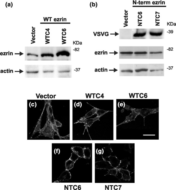
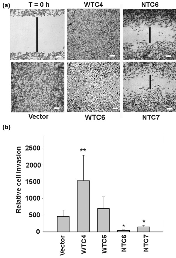
Methods: We examined the role of ezrin in metastasis using a highly metastatic murine mammary carcinoma cell line, namely AC2M2. Stable cell clones that overexpress wild-type ezrin or a dominant-negative amino-terminal domain of ezrin were selected. They were then tested for cell motility and invasion in vitro, and metastasis in a mouse in vivo tumour transplantation model.

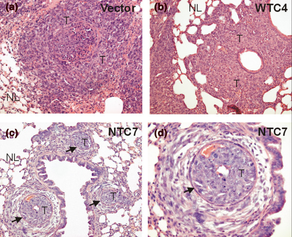
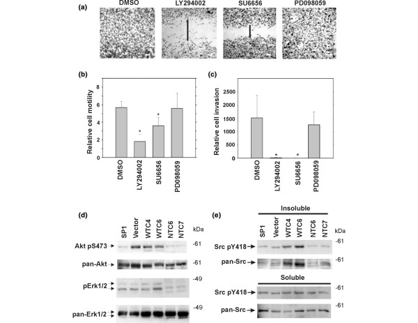
Results: Parental AC2M2 cells and cells overexpressing wild-type ezrin were transplanted into the mammary fat pad of syngeneic recipient mice; these animals subsequently developed lung metastases. In contrast, expression of the dominant-negative amino-terminal ezrin domain markedly inhibited lung metastasis. Consistent with this effect, we observed that the expression of amino-terminal ezrin caused strong membrane localization of cadherin, with increased cell-cell contact and a decrease in cell motility and invasion, whereas cells expressing wild-type ezrin exhibited strong cytoplasmic expression of cadherins and pseudopodia extensions. In addition, inhibitors of phosphatidylinositol 3-kinase and c-Src significantly blocked cell motility and invasion of AC2M2 cells expressing wild-type ezrin. We further found that overexpression of amino-terminal ezrin reduced levels of Akt pS473 and cytoskeletal-associated c-Src pY418 in AC2M2 cells, which contrasts with the high levels of phosphorylation of these proteins in cells expressing wild-type ezrin. Phosphorylated Erk1/2 was also reduced in amino-terminal ezrin expressing cells, although a mitogen-activated protein kinase kinase (MEK) inhibitor had no detectable effect on cell motility or invasion in this system.

Conclusion: Our findings indicate that ezrin is required for breast cancer metastasis, and that c-Src and phosphatidylinositol 3-kinase/Akt are effectors of ezrin in the cell motility and invasion stages of the metastatic process. Together, these results suggest that blocking ezrin function may represent a novel and effective strategy for preventing breast cancer metastasis.


Figures

Figure 1
Figure 1
Localization of cadherins in metastatic mammary carcinoma cells overexpressing wild-type and amino-terminal ezrin. Metastatic AC2M2 cells were transfected with empty pCB6 vector, or a vector encoding wild-type or amino-terminal ezrin, as described in the text. (a) Serial dilutions of the total cell extracts (0.6–20 μg) were subjected to reduced 10% SDS-PAGE and transferred to PVP membranes. The membranes were probed with anti-ezrin and anti-actin antibodies, followed by the appropriate peroxidase-conjugated secondary antibodies, and developed with chemiluminescence. Lanes from left to right contained 10 μg of the following cell extracts: pooled AC2M2 cells transfected with empty pCB6 vector, and two clones transfected with wild-type (WT) ezrin. WTC4 and WTC6 exhibited 4-fold and 8-fold overexpression, respectively, of ezrin compared with vector control cells. (b) Membranes were probed with anti-vesicular stomatitis virus glycoprotein (VSVG), anti-ezrin and anti-actin antibodies. Lanes contained 15 μg of cell extracts from pooled pCB6-transfected cells, and two clones transfected with amino-terminal ezrin. Clones NTC6 and NTC7 exhibited 1.6-fold and 4.6-fold amino-terminal ezrin expression, respectively, compared with endogenous ezrin, as determined by densitometric analysis of VSVG and ezrin blots normalized to actin. (c-g) The above cell lines were immunostained with anti-pan cadherin antibody, as described the text. Representative confocal microscope images are shown.
Figure 2
Figure 2
Expression of amino-terminal ezrin significantly reduces lung metastases of carcinoma cells. Metastatic AC2M2 cells transfected with empty pCB6 vector or a vector encoding wild-type ezrin or amino-terminal ezrin were injected (7.5 × 10³/mouse) into the mammary fat pad of syngeneic mice, and metastasis was assessed 6 weeks later, as described in the text. Images are shown from tissue sections from the lungs of mice injected with AC2M2 cells transfected with (a) empty pCB6 vector, (b) wild-type ezrin (WTC4), or (c,d) amino-terminal ezrin, stained with haematoxylin and eosin. Normal lung tissue is indicated by 'NL'. Tumour metastases are indicated by 'T'. Arrows indicate endothelial lining of vascular channels with tumour emboli. Original magnifications: panels a–c, 200×; panel d, 400×.
Figure 3
Figure 3
Expression of amino-terminal ezrin inhibits motility and invasion of carcinoma cells. (a) Transfected AC2M2 clones (see Fig. 1) were grown to confluence in 12-well NUNC tissue culture plates with 2 ml of 10% foetal bovine serum (FBS)/Dulbecco's modified Eagle medium (DMEM). Cultures were wounded by streaking cell monolayers with a 20 μl Eppendorf micropipette tip and monitored up to 24 hours, as described in the text. Representative fields photographed after 18 hours are shown. Vertical bars indicate the wound distance. Bar, 50 μm. (b) Transfected AC2M2 cells were subjected to an invasion assay, as described in the text. Cells (5 × 10⁴) were over-layered in 200 μl of 0.5% FBS/DMEM medium on the transwell membranes (8 μm pore size), with 0.5 ml of complete medium in the lower chamber. After 36–48 hours, cells were fixed and stained with modified haematoxylin, and cells invading through the membrane were counted using ImagePro software, as described in the text. The numbers of invading cells were normalized to empty pCB6 vector group in each experiment. The results from at least three independent experiments were pooled, and expressed as the mean relative cell invasion ± standard deviation. Asterisks indicate (**) a significant increase or (*) a significant reduction in cell invasion compared with vector control, using a two-sided Fisher's t-test. P = 0.03 for WTC4**; P = 0.009 for NTC6*; and P = 0.02 for NTC7*.
Figure 4
Figure 4
Role of phosphatidylinositol 3-kinase (PI3K) and c-Src in ezrin-mediated cell motility and invasion. (a) In a wound healing experiment with WTC4 cells, the PI3K inhibitor LY294002 (10 μmol/l), the c-Src inhibitor SU6656 (10 μmol/l), or the mitogen-activated protein kinase kinase (MEK) inhibitor (PD098059; 30 μmol/l), or the solvent DMSO (dimethyl sulfoxide; 10 μl/culture) was added to cultures, and wound closure was monitored up to 24 hours, as described for Fig. 3a. Representative fields photographed after 24 hours are shown. (b) The histogram shows pooled results from clones WTC4 and WTC6 in three independent wound healing experiments with the above inhibitors. Significant reduction in motility was observed in groups treated with LY294002 (P= 0.002) and SU6656 (P = 0.03). (c) WTC4 cells were set up in transwell cultures with PI3K, c-Src, or MEK inhibitors at the concentrations indicated above, and cell invasion was assessed after 36 hours. Results are expressed as the mean cell invasion ± standard deviation of at least three independent experiments. Single asterisk (*) indicates a specific reduction in invasion compared with DMSO-treated cells (LY294002, P = 0.02; SU6656, P = 0.02). (d) For analysis of Akt and Erk1/2 activation, transfected AC2M2 cell lines (see Fig. 1a) were serum starved overnight and cultured on fibronectin substratum (10 μg/ml) for 45 min. Cells were then lysed, and equal protein amounts of each cell lysate were subjected to 10% SDS-PAGE under reduced conditions. Proteins were transferred to PVP membranes, and western blotting was carried out with antibodies against Akt pS473, pan-Akt, Erk1/2 pT185/pY187 and pan-Erk1/2. (e) For c-Src analysis, cells were plated for 2 hours on fibronectin substratum and cell lysates of Triton X-100 soluble and insoluble (cytoskeletal-associated) fractions were prepared, as described in the text. Blots were probed with antibodies against c-Src pY418 and pan c-Src.

References

    1. Frame MC. Newest findings on the oldest oncogene; how activated Src does it. J Cell Sci. 2004;117:989–998. doi: 10.1242/jcs.01111. - DOI - PubMed
    1. Bretscher A, Edwards K, Fehon RG. ERM proteins and merlin: integrators at the cell cortex. Nat Rev Mol Cell Biol. 2002;3:586–599. doi: 10.1038/nrm882. - DOI - PubMed
    1. Gautreau A, Louvard D, Arpin M. ERM proteins and NF2 tumor suppressor: the Yin and Yang of cortical actin organization and cell growth signaling. Curr Opin Cell Biol. 2002;14:104–109. doi: 10.1016/S0955-0674(01)00300-3. - DOI - PubMed
    1. Pujuguet P, Del Maestro L, Gautreau A, Louvard D, Arpin M. Ezrin regulates E-cadherin-dependent adherens junction assembly through Rac1 activation. Mol Biol Cell. 2003;14:2181–2191. doi: 10.1091/mbc.E02-07-0410. - DOI - PMC - PubMed
    1. Gautreau A, Poullet P, Louvard D, Arpin M. Ezrin, a plasma membrane-microfilament linker, signals cell survival through the phosphatidylinositol 3-kinase/Akt pathway. Proc Natl Acad Sci USA. 1999;96:7300–7305. doi: 10.1073/pnas.96.13.7300. - DOI - PMC - PubMed

Publication types