

Overexpression of beta1-chain-containing laminins in capillary basement membranes of human breast cancer and its metastases
- PMID: 15987446
- PMCID: PMC1175051
- DOI: 10.1186/bcr1011
Overexpression of beta1-chain-containing laminins in capillary basement membranes of human breast cancer and its metastases
Abstract
Introduction: Laminins are the major components of vascular and parenchymal basement membranes. We previously documented a switch in the expression of vascular laminins containing the alpha4 chain from predominantly laminin-9 (alpha4beta2gamma1) to predominantly laminin-8 (alpha4beta1gamma1) during progression of human brain gliomas to high-grade glioblastoma multiforme. Here, differential expression of laminins was studied in blood vessels and ductal epithelium of the breast.
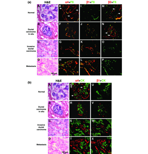
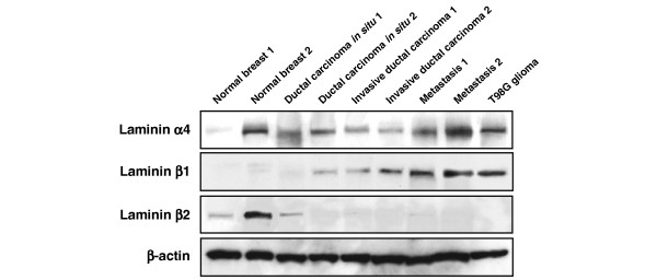
Method: In the present study the expressions of laminin isoforms alpha1-alpha5, beta1-beta3, gamma1, and gamma2 were examined during progression of breast cancer. Forty-five clinical samples of breast tissues including normal breast, ductal carcinomas in situ, invasive ductal carcinomas, and their metastases to the brain were compared using Western blot analysis and immunohistochemistry for various chains of laminin, in particular laminin-8 and laminin-9.
Results: Laminin alpha4 chain was observed in vascular basement membranes of most studied tissues, with the highest expression in metastases. At the same time, the expression of laminin beta2 chain (a constituent of laminin-9) was mostly seen in normal breast and carcinomas in situ but not in invasive carcinomas or metastases. In contrast, laminin beta1 chain (a constituent of laminin-8) was typically found in vessel walls of carcinomas and their metastases but not in those of normal breast. The expression of laminin-8 increased in a progression-dependent manner. A similar change was observed from laminin-11 (alpha5beta2gamma1) to laminin-10 (alpha5beta1gamma1) during breast tumor progression. Additionally, laminin-2 (alpha2beta1gamma1) appeared in vascular basement membranes of invasive carcinomas and metastases. Chains of laminin-5 (alpha3beta3gamma2) were expressed in the ductal epithelium basement membranes of the breast and diminished with tumor progression.
Conclusion: These results suggest that laminin-2, laminin-8, and laminin-10 are important components of tumor microvessels and may associate with breast tumor progression. Angiogenic switch from laminin-9 and laminin-11 to laminin-8 and laminin-10 first occurs in carcinomas in situ and becomes more pronounced with progression of carcinomas to the invasive stage. Similar to high-grade brain gliomas, the expression of laminin-8 (and laminin-10) in breast cancer tissue may be a predictive factor for tumor neovascularization and invasion.
Figures
Comment in
-
Laminin isoform expression in breast tumors.Breast Cancer Res. 2005;7(4):166-7. doi: 10.1186/bcr1270. Epub 2005 May 24. Breast Cancer Res. 2005. PMID: 15987470 Free PMC article.
References
-
- Nakhlis F, Morrow M. Ductal carcinoma in situ. Surg Clin North Am. 2003;83:821–839. - PubMed
-
- Weidner N, Folkman J, Pozza F, Bevilacqua P, Allred EN, Moore DH, Meli S, Gasparini G. Tumor angiogenesis: a new significant and independent prognostic indicator in early-stage breast carcinoma. J Natl Cancer Inst. 1992;84:1875–1887. - PubMed
Publication types
MeSH terms
Substances
Grants and funding
LinkOut - more resources
Full Text Sources
Other Literature Sources
Medical
