. 2005;7(4):R422-35.
doi: 10.1186/bcr1019. Epub 2005 Apr 4.

Mechanisms underlying the growth inhibitory effects of the cyclo-oxygenase-2 inhibitor celecoxib in human breast cancer cells

Affiliations

Mechanisms underlying the growth inhibitory effects of the cyclo-oxygenase-2 inhibitor celecoxib in human breast cancer cells

Gargi D Basu et al. Breast Cancer Res. 2005.

Abstract

Introduction: Inhibitors of cyclo-oxygenase (COX)-2 are being extensively studied as anticancer agents. In the present study we evaluated the mechanisms by which a highly selective COX-2 inhibitor, celecoxib, affects tumor growth of two differentially invasive human breast cancer cell lines.

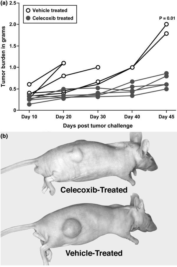
Methods: MDA-MB-231 (highly invasive) and MDA-MB-468 (moderately invasive) cell lines were treated with varying concentrations of celecoxib in vitro, and the effects of this agent on cell growth and angiogenesis were monitored by evaluating cell proliferation, apoptosis, cell cycle arrest, and vasculogenic mimicry. The in vitro results of MDA-MB-231 cell line were further confirmed in vivo in a mouse xenograft model.

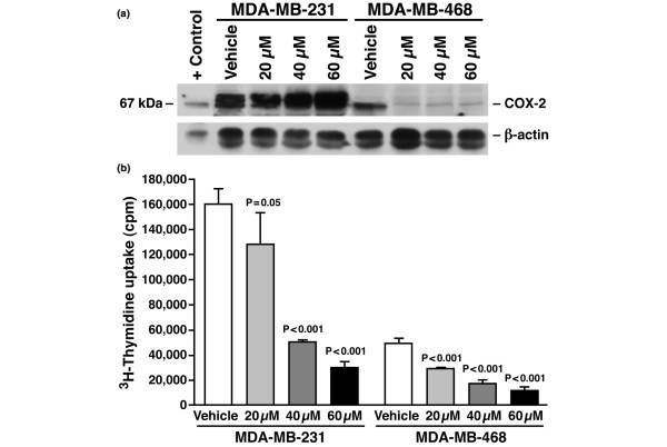
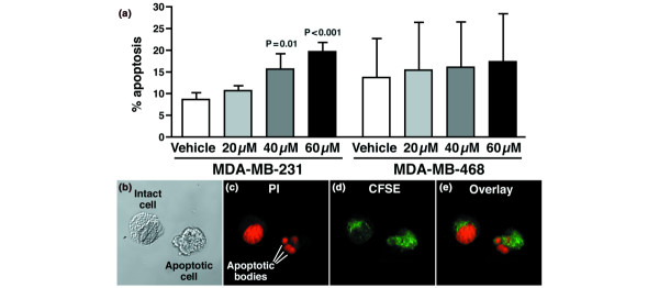
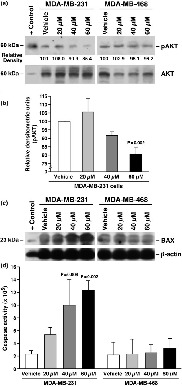
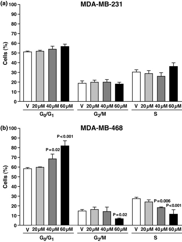
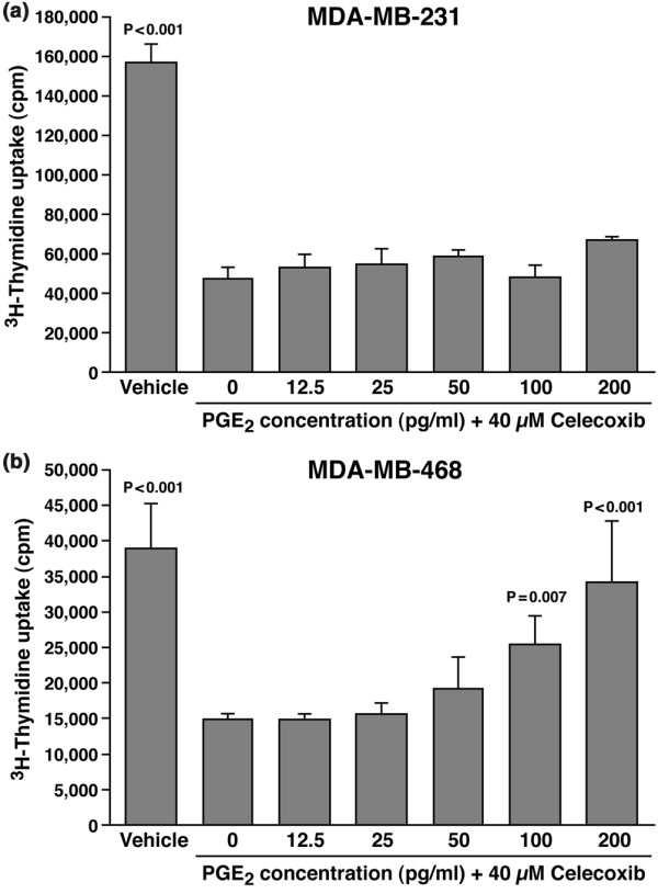
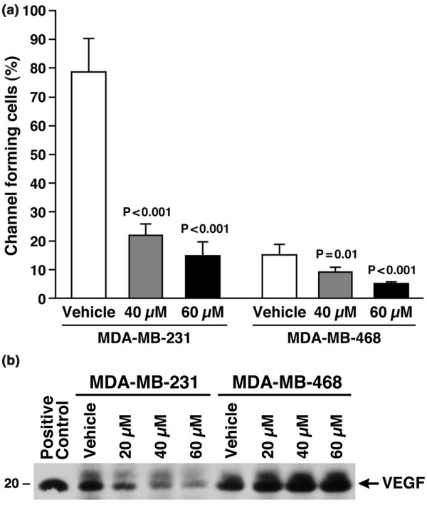
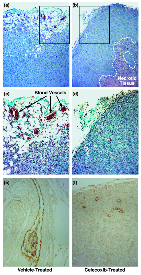
Results: The highly invasive MDA-MB-231 cells express higher levels of COX-2 than do the less invasive MDA-MB-468 cells. Celecoxib treatment inhibited COX-2 activity, indicated by prostaglandin E2 secretion, and caused significant growth arrest in both breast cancer cell lines. In the highly invasive MDA-MB-231 cells, the mechanism of celecoxib-induced growth arrest was by induction of apoptosis, associated with reduced activation of protein kinase B/Akt, and subsequent activation of caspases 3 and 7. In the less invasive MDA-MB-468 cells, growth arrest was a consequence of cell cycle arrest at the G0/G1 checkpoint. Celecoxib-induced growth inhibition was reversed by addition of exogenous prostaglandin E2 in MDA-MB-468 cells but not in MDA-MB-231 cells. Furthermore, MDA-MB-468 cells formed significantly fewer extracellular matrix associated microvascular channels in vitro than did the high COX-2 expressing MDA-MB-231 cells. Celecoxib treatment not only inhibited cell growth and vascular channel formation but also reduced vascular endothelial growth factor levels. The in vitro findings corroborated in vivo data from a mouse xenograft model in which daily administration of celecoxib significantly reduced tumor growth of MDA-MB-231 cells, which was associated with reduced vascularization and increased necrosis in the tumor mass.

Conclusion: The disparate molecular mechanisms of celecoxib-induced growth inhibition in human breast cancer cells depends upon the level of COX-2 expression and the invasive potential of the cell lines examined. Data suggest a role for COX-2 not only in the growth of cancer cells but also in activating the angiogenic pathway through regulating levels of vascular endothelial growth factor.


Figures

Figure 1
Figure 1
Celecoxib regulates COX-2 levels and causes growth arrest in human breast cancer cells. (a) Cyclo-oxygenase (COX)-2 is expressed in both MDA-MB-231 and MDA-MB-468 cell lines. Western blot analysis of vehicle and celecoxib (20–60 μmol/l) treated cells. SDS-PAGE electrophoresis was performed using a 10% resolving gel. Protein was loaded at 100 μg per lane. Lipopolysaccharide/phorbol 12-myristate 13-acetate treated whole cell lysate from RAW264.7 cell line was used as positive control. Gels were blotted and probed with COX-2 monoclonal antibody. Both cell lines expressed COX-2. MDA-MB-231 cells expressed higher levels of COX-2 than did MDA-MB-468 cells. With drug treatment, COX-2 protein level did not change in the MDA-MB-231 cells, but there was reduction in the level of COX-2 protein in the MDA-MB-468 cells after treatment. β-Actin blot is included to confirm equal loading. These experiments were repeated three times with similar results. (b) Celecoxib induced dose-dependent inhibition of proliferation of breast cancer cell lines. Cells were incubated for 4 days with vehicle or celecoxib, and [3H]thymidine was added 24 hours before harvest. After washing off excess thymidine, cells were lysed with 5% Triton X-100, and incorporated thymidine was evaluated. Celecoxib treatment caused significant dose-dependent growth inhibition in both human breast cancer cell lines. Mean values of three experiments ± standard deviation is shown. P values represent significant differences between vehicle control and celecoxib treatment.
Figure 2
Figure 2
Celecoxib induces apoptosis in MDA-MB-231 cells. (a) Flow cytometric analysis of vehicle-treated and celecoxib-treated cells stained with annexin V and propidium iodide (PI) was done 48 hours after treatment. The population shown in the figure is total apoptotic cells, which includes early and late apoptosis. Significant induction of apoptosis was observed in the MDA-MB-231 cells at 40 and 60 μmol/l concentrations of celecoxib. Apoptosis was not induced in MDA-MB-468 cells. Mean values of three experiments ± standard deviation is shown. P values represent significant differences between vehicle control and celecoxib treatment. (b–e) Celecoxib induces formation of apoptotic bodies in MDA-MB-231 cells. Shown are confocal images of MDA-MB-231 cells subjected to 48 hours of celecoxib (60 μmol/l) treatment. Cells were stained with CFSE (panel d) and then fixed in 95% ethanol and stained with PI (panel c). Cells were visualized in a confocal microscope (Carl Zeiss Inc.) using excitation wavelengths of 488 nm (for CFSE) and 543 nm (for PI). Loss of integrity of nuclear envelope and formation of peripheral, sharply delineated masses of condensed chromatin or apoptotic bodies are visualized. Panel b represents phase contrast images of the cells and panel e represents colocalization of CFSE and PI. Images were taken 200×.
Figure 3
Figure 3
Celecoxib induced down-regulation of pAkt, increase in Bax, and caspase 3/7 in MDA-MB-231 cells. (a) Total Akt and phosphorylated Akt (pAkt). Western blot analysis of cell lysates prepared from vehicle and celecoxib (20–60 μmol/l) treated cells. SDS-PAGE electrophoresis was performed using 10% resolving gel. Protein was loaded at 100 μg per lane and the protein of interest was detected using specific antibodies. Celecoxib treatment at 40 and 60 μmol/l caused decreases in the levels of pAkt in MDA-MB-231 cells, with no change in MDA-MB-468 cells. Numbers below each lane represents percentage of protein expression compared with vehicle-treated cell lysate, which was set to equivalent to 100%, as determined by densitometric analysis. Control cell extracts from Jurkats were used as positive control for Akt and pAkt. (b) Average densitometric values of three separate experiments showing reduction in pAkt with celecoxib treatment. There was a significant decrease (P = 0.002) in the levels of pAkt with 60 μmol/l celecoxib treatment. (c) Western blot analysis of BAX. Increased expression of BAX protein was observed with increasing concentrations of celecoxib in MDA-MB-231 cells but not in MDA-MB-468 cells. The experiment was repeated three times with similar results. A β-actin blot is included to show equal loading. (d) Spectrofluorometric analysis of lysates prepared from vehicle and celecoxib (20–60 μmol/l) treated cells at 48 hours. Activity of caspases 3 and 7 was monitored by enzymatic cleavage using a fluorescence microplate reader with excitation at 485 ± 10 nm and emission detection at 530 ± 12.5 nm. In MDA-MB-231 cells, activities of caspases 3 and 7 were increased significantly at 40 μmol/l and 60 μmol/l drug concentrations. No increase in caspase activity was evident in the MDA-MB-468 cells. Mean values from three experiments ± standard deviation is shown. P values represent significant differences between vehicle control and celecoxib treatment.
Figure 4
Figure 4
Celecoxib causes cell cycle arrest in MDA-MB-468 cells. (a,b) Flow cytometric analysis of cells subjected to treatment with vehicle or celecoxib (20–60 μmol/l) for 48 hours. Cells were fixed and permeabilized with 95% ethanol, stained with propidium iodide, and analyzed by flow cytometry. Celecoxib induced growth arrest at the G0/G1 cell cycle checkpoint in MDA-MB-468 cells (panel b) with no cell cycle arrest in the MDA-MB-231 cells (panel a). Mean values for three experiments ± standard deviation of the mean is shown. P values represent significant difference between vehicle control and celecoxib treatment. Experiments were repeated three times, with similar results.
Figure 5
Figure 5
Growth inhibition of MDA-MB-468 cells was abrogated by exogenous prostaglandin (PG)E2 addition. [3H]thymidine uptake assay was done to determine proliferation of (a) MDA-MB-231 and (b) MDA-MB-468 cells treated with 40 μmol/l celecoxib with or without varying amounts of exogenous PGE2 (12.5–200 pg/ml). Cells were harvested after 96 hours in culture. In MDA-MB-231 cells, growth inhibition induced by 40 μmol/l celecoxib could not be restored by addition of exogenous PGE2; however, addition of 200 pg/ml PGE2 completely reversed the growth inhibition induced by 40 μmol/l celecoxib in the less invasive MDA-MB-468 cells. Average values of three experiments ± standard deviation is shown. P values represent significant differences between vehicle control and celecoxib treatment.
Figure 6
Figure 6
Celecoxib treatment causes reduction in microvascular channel formation by regulating VEGF levels. (a) The percentage of cells forming channels was much greater in MDA-MB-231 cells than in MDA-MB-468 cells. In both cells, treatment with 40 and 60 μmol/l celecoxib caused significant reduction in the number of channels. P values represent significant differences between vehicle control and celecoxib treatment. (b) Western blot analysis of cell lysates prepared from vehicle and celecoxib (20–60 μmol/l) treated cells. SDS-PAGE electrophoresis was performed using 15% resolving gel. Protein was loaded at 100 μg per lane and the protein was detected using vascular endothelial growth factor (VEGF) antibody. Celecoxib treatment decreased VEGF levels in MDA-MB-231 cells in a dose-dependent manner.
Figure 7
Figure 7
Celecoxib treatment reduced MDA-MB-231 tumor growth in nude mice. (a) Five mice per group were treated with either celecoxib (25 mg/kg body weight) or vehicle (dimethyl sulfoxide) and the mice were killed 45 days after the tumor cells were inoculated. Tumor growth was monitored by weekly examination using digital calipers, and tumor weight was calculated using to the following equation [23]: tumor weight (g) = (length (cm) × width (cm)2) × 0.5. Three mice from the vehicle-treated group had to be killed early because of the aggressive nature of the tumor. The other two mice in the vehicle-treated group had significantly greater tumor burden (P = 0.01) than did the five mice in celecoxib-treated group. (b) A representative mouse from each treatment group is illustrated; lower tumor mass Is evident in the treated animal as compared with the vehicle control.
Figure 8
Figure 8
In vivo inhibition of angiogenesis and increase in necrosis with celecoxib treatment. Vascularity of tumor implants was histologically evaluated by Masson's trichrome and factor VIII related antigen staining. Shown is evidence of central necrosis and decreased number of blood vessels in (b) a section of celecoxib-treated tumors relative to (a) a section obtained from a vehicle-treated animal (magnification 50×). Greater magnification (100×) of (c) panel a and (d) panel b are shown in the next two panels. Arrows in panel c point to blood vessels. Endothelial cells lining the blood vessels stained positively for factor VIII related antigen and showed larger blood vessels in the (e) vehicle-treated than in the (f) celecoxib-treated samples (magnification 100×).

References

    1. Harris RE, Chlebowski RT, Jackson RD, Frid DJ, Ascenseo JL, Anderson G, Loar A, Rodabough RJ, White E, McTiernan A. Breast cancer and nonsteroidal anti-inflammatory drugs: prospective results from the Women's Health Initiative. Cancer Res. 2003;63:6096–6101. - PubMed
    1. Lee PP, Ip MM. Regulation of proliferation of rat mammary tumor cells by inhibitors of cyclooxygenase and lipoxygenase. Prostaglandins Leukot Essent Fatty Acids. 1992;45:21–31. doi: 10.1016/0952-3278(92)90098-4. - DOI - PubMed
    1. Robertson FM, Parrett ML, Joarder FS, Ross M, Abou-Issa HM, Alshafie G, Harris RE. Ibuprofen-induced inhibition of cyclooxygenase isoform gene expression and regression of rat mammary carcinomas. Cancer Lett. 1998;122:165–175. doi: 10.1016/S0304-3835(97)00387-X. - DOI - PubMed
    1. Alshafie GA, Harris RE, Robertson FM, Parrett ML, Ross M, Abou-Issa H. Comparative chemopreventive activity of ibuprofen and N-(4-hydroxyphenyl) retinamide against the development and growth of rat mammary adenocarcinomas. Anticancer Res. 1999;19:3031–3036. - PubMed
    1. Zhao Y, Agarwal VR, Mendelson CR, Simpson ER. Estrogen biosynthesis proximal to a breast tumor is stimulated by PGE2 via cyclic AMP, leading to activation of promoter II of the CYP19 (aromatase) gene. Endocrinology. 1996;137:5739–5742. doi: 10.1210/en.137.12.5739. - DOI - PubMed

Publication types