Comparative Study
. 2005;7(4):R769-76.
doi: 10.1186/ar1728. Epub 2005 Apr 4.

A monoclonal antibody against kininogen reduces inflammation in the HLA-B27 transgenic rat

Affiliations
Comparative Study

A monoclonal antibody against kininogen reduces inflammation in the HLA-B27 transgenic rat

James C Keith Jr et al. Arthritis Res Ther. 2005.

Abstract

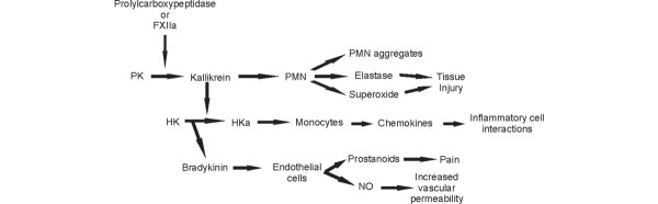
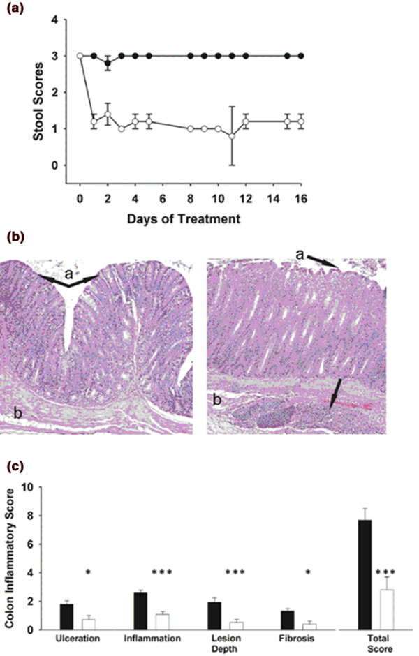
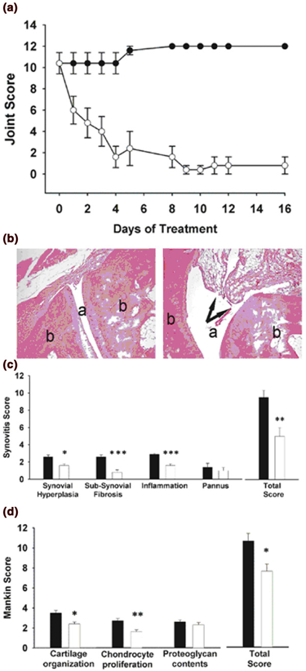
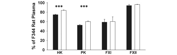
The human leukocyte antigen B27 (HLA-B27) transgenic rat is a model of human inflammatory bowel disease, rheumatoid arthritis and psoriasis. Studies of chronic inflammation in other rat models have demonstrated activation of the kallikrein-kinin system as well as modulation by a plasma kallikrein inhibitor initiated before the onset of clinicopathologic changes or a deficiency in high-molecular-mass kininogen. Here we study the effects of monoclonal antibody C11C1, an antibody against high-molecular-mass kininogen that inhibits the binding of high-molecular-mass kininogen to leukocytes and endothelial cells in the HLA-B27 rat, which was administered after the onset of the inflammatory changes. Thrice-weekly intraperitoneal injections of monoclonal antibody C11C1 or isotype IgG1 were given to male 23-week-old rats for 16 days. Stool character as a measure of intestinal inflammation, and the rear limbs for clinical signs of arthritis (tarsal joint swelling and erythema) were scored daily. The animals were killed and the histology sections were assigned a numerical score for colonic inflammation, synovitis, and cartilage damage. Administration of monoclonal C11C1 rapidly decreased the clinical scores of pre-existing inflammatory bowel disease (P < 0.005) and arthritis (P < 0.001). Histological analyses confirmed significant reductions in colonic lesions (P = 0.004) and synovitis (P = 0.009). Decreased concentrations of plasma prekallikrein and high-molecular-mass kininogen were found, providing evidence of activation of the kallikrein-kinin system. The levels of these biomarkers were reversed by monoclonal antibody C11C1, which may have therapeutic potential in human inflammatory bowel disease and arthritis.


Figures

Figure 1
Figure 1
Kallikrein–kinin system (KKS). The KKS is initiated by factor XIIa (FXIIa) or prolylcarboxypeptidase on the endothelial cell and leukocyte (polymorphonuclear cell (PMN)) surface, generating the enzyme kallikrein, which in turn cleaves high-molecular-mass kininogen (HK) to yield bradykinin (BK) and cleaved high-molecular-mass kininogen (HKa). Kallikrein is chemotactic, aggregates neutrophils, and stimulates the release of elastase and superoxide (potent inducers of tissue injury). BK stimulates vasodilation, mediates pain through the release of prostaglandins, and stimulates vascular permeability through the generation of nitrous oxide (NO). PK, prekallikrein.
Figure 2
Figure 2
Effect of mAb C11C1 on HLA-B27 transgenic rats colonic inflammation. (a) Effects of monoclonal antibody (mAb) C11C1 on diarrhea in human leukocyte antigen B27 (HLA-B27) rats. Stool score was determined five times a week (normal stool = 1, soft stool = 2, watery stool = 3). mAb C11C1 (1.9 mg/kg) was administered three times a week for 16 days. The control group received murine isotype IgG1 (6 mg/kg) three times a week for 16 days. All stool scores are significantly different between the two groups for each corresponding day (P < 0.005) except for day 11 (P = 0.03). Data are shown as means ± SEM. Filled circles, IgG1-treated group; open circles, mAb C11C1-treated group. (b) Effects of mAb C11C1 on colonic mucosa in HLA-B27 rats. Photomicrographs of representative sections of colon from C11C1-treated (left) and IgG-treated (right) HLA-B27 transgenic rats. Note the extensive inflammatory cell infiltrates within the mucosa (a) and submucosa (b) with loss of villus formation on the mucosal surface indicated by the arrow (a) in the IgG group (right) compared with the C11C1 group (left). The branched arrow (left) points to the villus formation normally present in the colon (mAb C11C1-treated group). Hematoxylin and eosin stain; original magnification × 100. (c) Effects of mAb C11C1 on colonic inflammatory changes in HLA-B27 rats. mAb C11C1 decreased inflammatory changes in the colonic sections as evaluated by ulceration (P = 0.02), inflammation (P < 0.001), depth of lesion (P = 0.004), and degree of fibrosis replacement (P = 0.01) compared with IgG1 administration. Treatment with mAb C11C1 (open bars) significantly decreased the extent and intensity of the total colonic inflammatory score (P = 0.004). Data are shown as means ± SEM. *P < 0.05; ***P < 0.005.
Figure 3
Figure 3
Effect of mAb C11C1 on HLA-B27 transgenic rat inflammatory arthritis. (a) Effects of monoclonal antibody (mAb) C11C1 on clinical signs of arthritis in human leukocyte antigen B27 (HLA-B27) rats. mAb C11C1 was administered at the same dose and frequency as in Fig. 2a. Mean joint score was determined daily, except at weekends. All joint scores are significantly different between the two groups for each corresponding day (P < 0.001) except for days 1 (P > 0.03), 2 (P = 0.01) and 3 (P = 0.006). Data are shown as means ± SEM. Filled circles, IgG1-treated group; open circles, mAb C11C1-treated group. (b) Effects of mAb C11C1 on joint histology in HLA-B27 rats. Photomicrographs of representative sections of tarsal joints from C11C1-treated (left) and IgG-treated (right) HLA-B27 transgenic rats. Note the clear joint space (a) and normal appearance of bone (b) in the mAb C11C1-treated group (left) compared with the inflamed villus formation (arrows) occupying the synovial space (a) in the IgG-treated group (right). Hematoxylin and eosin stain; original magnification × 100. (c) Effects of mAb C11C1 on synovial inflammatory changes in HLA-B27 rats. Treatment with mAb C11C1 (open bars) decreased synovial proliferation (hyperplasia) (P = 0.01), subsynovial fibrosis (fibroplasia) (P = 0.001), and degree of inflammation (P < 0.001), but not pannus formation. The total score of the control IgG1 of 9.6 ± 1.0 was reduced by mAb C11C1 to an inflammatory score of 5.0 ± 1.0 (P = 0.009). Data are shown as means ± SEM. *P < 0.05; **P < 0.01; ***P < 0.005. (d) Effects of mAb C11C1 on cartilage and bone inflammatory changes in HLA-B27 rats. mAb C11C1 (open bars) significantly improved (decreased the Mankin score of) the cartilage organization (P = 0.01) and the altered chondrocyte proliferation (P = 0.008). The proteoglycan cartilage contents (Safranin O/Fast Green staining) were similar in both experimental groups (P > 0.05) and the tidemark integrity was preserved (data not shown). The total Mankin score was significantly decreased in the mAb C11C1-treated group (P = 0.02). Data are shown as means ± SEM. *P < 0.05; **P < 0.01; ***P < 0.005.
Figure 4
Figure 4
Kallikrein–kinin system (KKS) assays. Plasma KKS protein concentrations in the human leukocyte antigen B27 transgenic rats treated with control monoclonal antibody IgG (filled bars) or monoclonal antibody C11C1 (open bars) at day 16 of the experimental protocol. Values were compared with a pool of normal Fischer 344 rat plasma. Both high-molecular-mass kininogen (HK) and prekallikrein (PK) were significantly decreased in the IgG1-treated group and were closer to normal in the C11C1-treated group. Both experimental groups showed decreased factor XI (FXI) with no significant differences between them. There were no significant changes between any groups in factor XII (FXII). ***P < 0.005.

References

    1. Taurog JD, Richardson JA, Croft JT, Simmons WA, Zhou M, Fernandez-Sueiro JL, Balish E, Hammer RE. The germfree state prevents development of gut and joint inflammatory disease in HLA-B27 transgenic rats. J Exp Med. 1994;180:2359–2364. doi: 10.1084/jem.180.6.2359. - DOI - PMC - PubMed
    1. Hammer RE, Maika SD, Richardson JA, Tang JP, Taurog JD. Spontaneous inflammatory disease in7 transgenic rats expressing HLA-B27 and human beta 2m: an animal model of HLA-B27-associated human disorders. Cell. 1990;63:1099–1112. doi: 10.1016/0092-8674(90)90512-D. - DOI - PubMed
    1. Sartor RB, Rath HC, Lichtman SN, van Tol EA. Animal models of intestinal and joint inflammation. Baillieres Clin Rheumatol. 1996;10:55–76. - PubMed
    1. Breban M. HLA-B27 transgenic rats model. Ann Med Interne (Paris) 1998;149:139–141. - PubMed
    1. Joseph K, Shibayama Y, Ghebrehiwet B, Kaplan AP. Factor XII-dependent contact activation on endothelial cells and binding proteins gC1qR and cytokeratin 1. Thromb Haemost. 2001;85:119–124. - PubMed

Publication types

MeSH terms