. 2005 Jun 29;2(1):12.
doi: 10.1186/1742-6413-2-12.

Fine-needle aspiration of the thyroid: an overview

Affiliations

Fine-needle aspiration of the thyroid: an overview

Gia-Khanh Nguyen et al. Cytojournal. .

Abstract

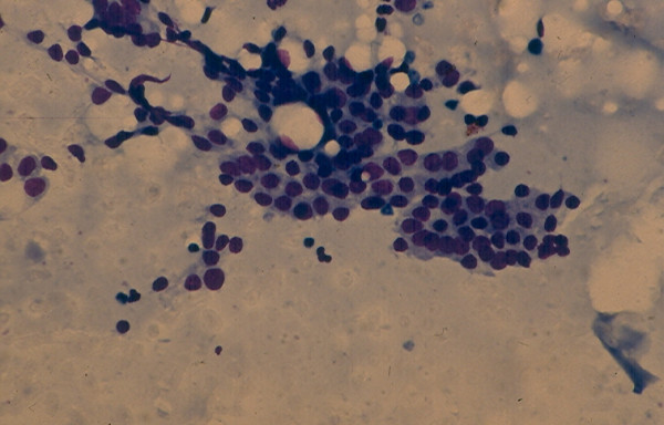
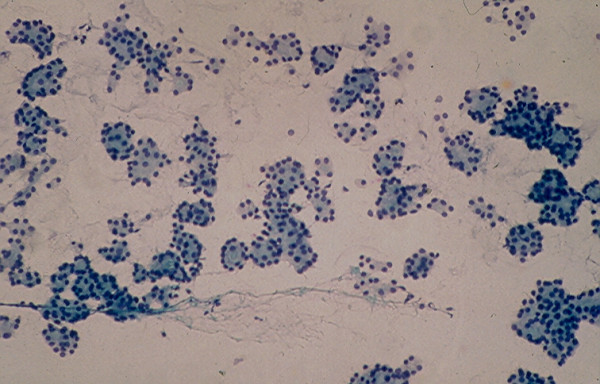
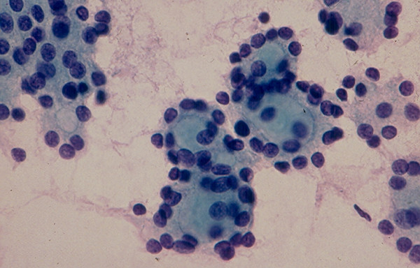
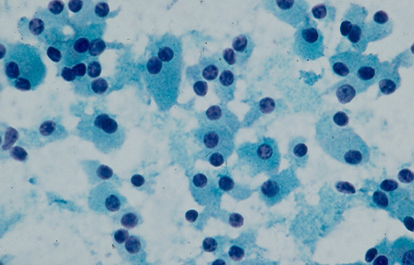
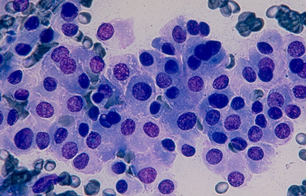
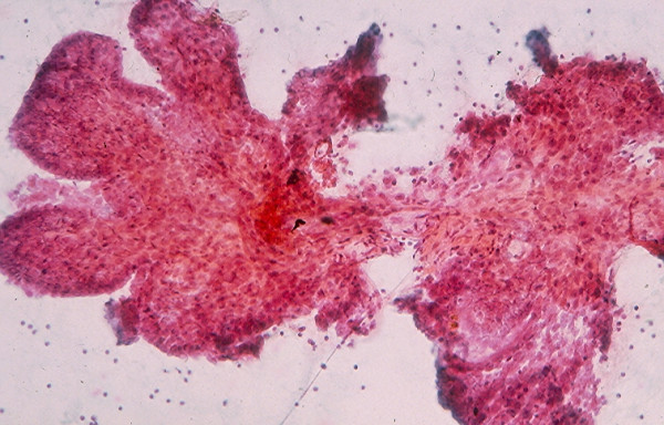
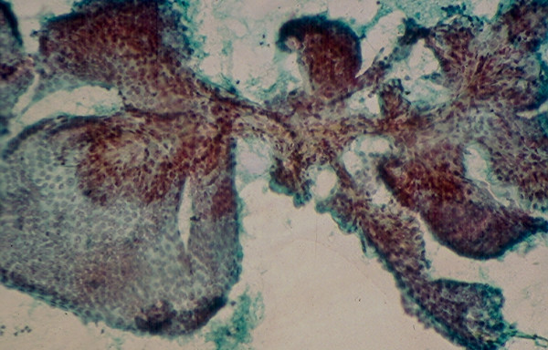
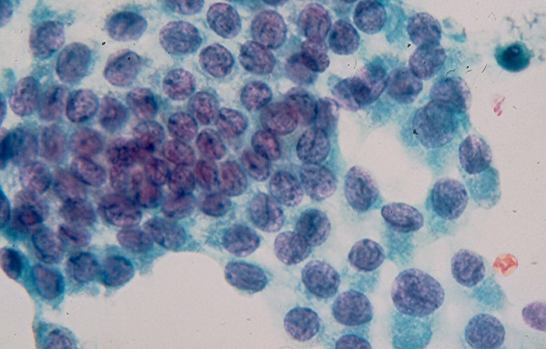
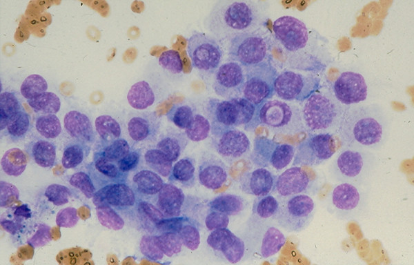
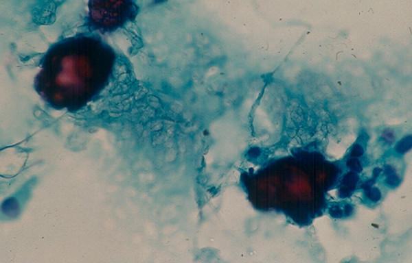
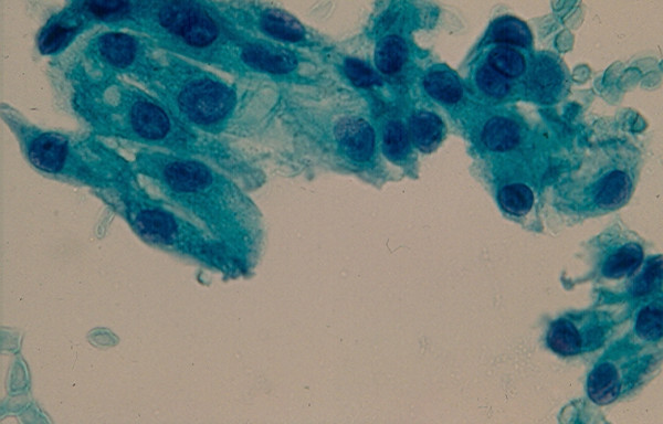
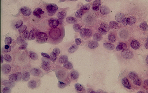
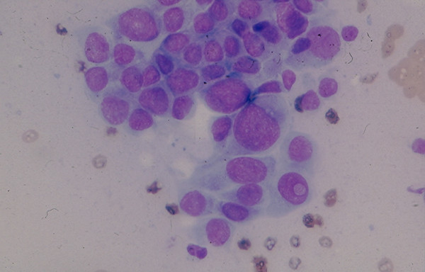
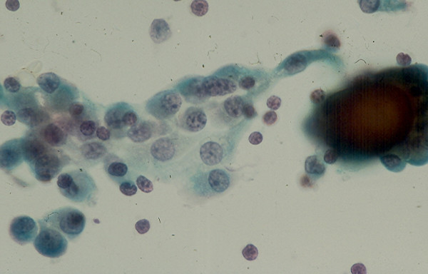
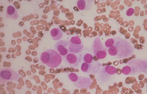
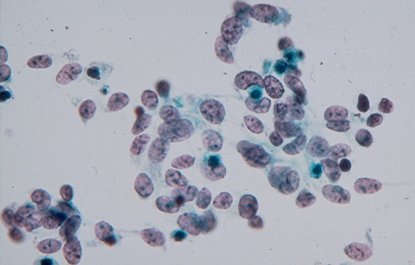
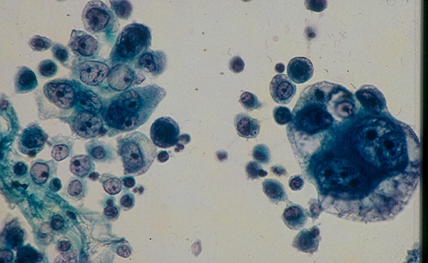
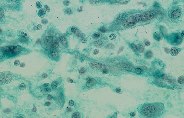
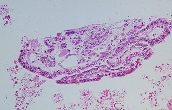
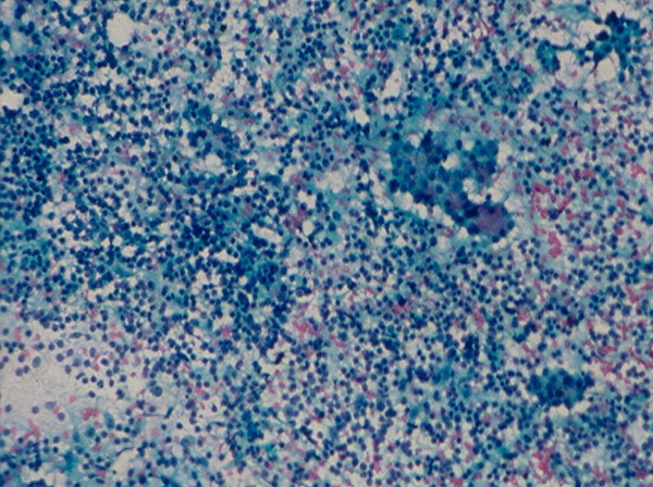
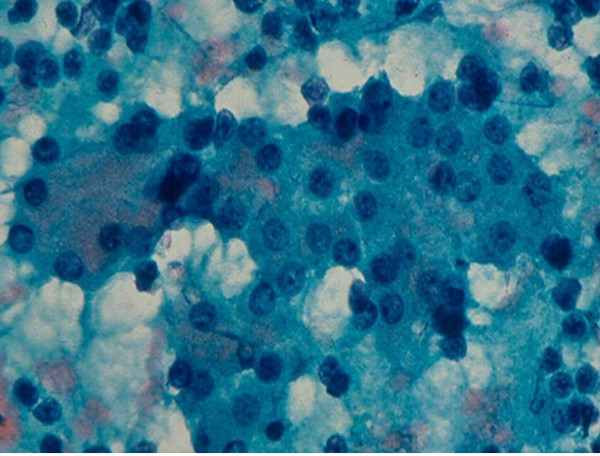
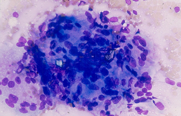
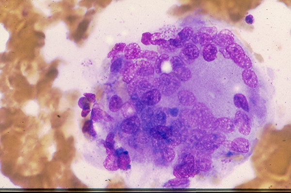
Thyroid nodules (TN) are a common clinical problem. Fine needle aspiration (FNA) of the thyroid now is practiced worldwide and proves to be the most economical and reliable diagnostic procedure to identify TNs that need surgical excision and TNs that can be managed conservatively. The key for the success of thyroid FNA consists of an adequate or representative cell sample and the expertise in thyroid cytology. The FNA cytologic manifestations of TNs may be classified into seven working cytodiagnostic groups consisting of a few heterogenous lesions each to facilitate the differential diagnosis. Recent application of diagnostic molecular techniques to aspirated thyroid cells proved to be useful in separating benign from malignant TNs in several cases of indeterminate lesions.


Figures

Figure 1
Figure 1
Thick, deep blue colloid material with cracking pattern in FNA of a benign colloid nodule (Diff-Quik stain, × 250).
Figure 2
Figure 2
Thick, deep blue colloid material with bubble pattern in FNA of a benign colloid nodule (Diff-Quik stain, × 250) view).
Figure 3
Figure 3
A monolayered sheet of benign follicular epithelial cells with honeycomb pattern in FNA of a benign colloid nodule (Diff-Quik stain, × 400).
Figure 4
Figure 4
Cellular microfollicular lesion showing in FNA cells with round nuclei arranged in acini and small monolayered sheet (Papanicolaou stain, 4 × 160 and 5 × 400).
Figure 5
Figure 5
Cellular microfollicular lesion showing in FNA cells with round nuclei arranged in acini (Papanicolaou stain, 4 × 160 and 5 × 400).
Figure 6
Figure 6
Hurthle cells with abundant, granular cytoplasm and round, central or eccentrically located nuclei in FNA of a Hurthle cell lesion (Papanicolaou stain, × 400).
Figure 7
Figure 7
Hurthle cells in loose, monolayered sheet and singly in FNA of a Hurthle cell lesion (Diff-Quik stain, × 400).
Figure 8
Figure 8
Thick branching papillary tissue fragment with fibrovascular core in FNA of a conventional papillary carcinoma (Papanicolaou stain, × 100).
Figure 9
Figure 9
Thin branching papillary tissue fragment with fibrovascular core in FNA of a conventional papillary carcinoma (Papanicolaou stain, × 100).
Figure 10
Figure 10
A sheet of tumor cells showing focal nuclear crowding with several cells displaying nuclear grooves in FNA of a conventional papillary carcinoma (Papanicolaou stain, × 400).
Figure 11
Figure 11
A loose sheet of tumor cells showing minimal nuclear crowding and two cells with intranuclear cytoplasmic inclusions in FNA of a conventional papillary carcinoma (Diff-Quik stain, × 400).
Figure 12
Figure 12
Two psammoma bodies in a smear showing a small amount of colloid material. A small aggregate of poorly preserved follicular cells is seen beside one psammoma body (Papanicolaou stain, × 400).
Figure 13
Figure 13
A loose cluster of metaplastic squamous cells seen in FNA of a conventional papillary carcinoma (Papanicolaou stain, × 400).
Figure 14
Figure 14
Papillary carcinoma, microfollicular variant showing in FNA cells in acinar arrangement. A tumor cell with an intranuclear cytoplasmic inclusion is noted (Papanicolaou stain, × 400).
Figure 15
Figure 15
Papillary carcinoma, tall-cell variant showing in FNA a sheet of pleomorphic cells with some cells with elongated configuration and cytoplasmic tails. A tumor cell with intranuclear cytoplasmic inclusion is present (Diff-Quik stain, × 400).
Figure 16
Figure 16
Papillary carcinoma, diffuse sclerosing type showing in FNA a sheet of metaplastic squamous cells, scattered lymphocytes and a psammoma body (Papanicolaou stain, × 400).
Figure 17
Figure 17
Medullary carcinoma showing in FNA dyshesive plasmacytoid tumor cells with eccentrically located round nuclei and intracytoplasmic azurophil granules (Diff-Quik stain, × 400).
Figure 18
Figure 18
Medullary carcinoma showing in FNA loosely clustered spindle-shaped tumor cells with scanty, ill-defined cytoplasm (Papanicolaou stain, × 400).
Figure 19
Figure 19
A fragment of orange and granular amyloid material seen in FNA of a thyroid medullary carcinoma (Papanicolaou stain, × 400).
Figure 20
Figure 20
Anaplastic carcinoma, giant-cell type showing in FNA single and clustered large, bizarre malignant cells with pleomorphic nuclei and prominent nucleoli (Papanicolaou stain, × 400).
Figure 21
Figure 21
Anaplastic carcinoma, spindle-cell type showing in FNA dyshesive spindle- shaped malignant cells with scant, ill-defined cytoplasm (Papanicolaou stain, × 400).
Figure 22
Figure 22
Papillary tissue fragments with thin fibrovascular cores covered with epithelial cells displaying nuclear crowding and occasional intranuclear cytoplasmic inclusions seen in a cell block section prepared from the needle aspirate of a papillary carcinoma with hemorrhagic cystic degenerative change (hematoxylin and eosin stain, × 250).
Figure 23
Figure 23
Section from a cell block prepared from the needle aspirate of a benign colloid nodule with hemorrhagic cystic degenerative change showing papillary tissue fragments covered with epithelium displaying no nuclear changes characteristic for a papillary carcinoma (hematoxylin and eosin stain, × 250).
Figure 24
Figure 24
Hashimoto thyroiditis showing in FNA numerous lymphoid cells admixed with a sheet of follicular epithelial cells (Papanicolaou stain, × 100).
Figure 25
Figure 25
A sheet of follicular epithelial cells with oncocytic change admixed with benign lymphoid cells seen in FNA of a Hashimoto thyroiditis (Papanicolaou stain, × 400).
Figure 26
Figure 26
A syncytial cluster of epithelioid cells with carrot-shaped nuclei seen in FNA of a subacute thyroiditis (Diff-Quik stain, × 400).
Figure 27
Figure 27
A large multinucleate giant cell present in FNA of a subacute thyroiditis (Diff-Quik stain, × 400).

References

    1. Martin HE, Ellis EB. Biopsy by needle puncture and aspiration. Am Surg. 1930;92:169–181. - PMC - PubMed
    1. Stewart FW. The diagnosis of tumors by aspiration. Am J Pathol. 1933;9:801–812. - PMC - PubMed
    1. Frazell EL, Foote FW. Papillary carcinoma of the thyroid: a review of 25 years of experience. Cancer. 1958;11:895–922. - PubMed
    1. Soderstrom N. Puncture of goiters for aspiration biopsy. A primary report. Acta Med Scand. 1952;144:237–244. - PubMed
    1. Eihorn J, Franzen S. Thin needle biopsy in the diagnosis of thyroid disease. Acta Radiol. 1962;58:321–336.