. 2005 Jun 29:3:6.
doi: 10.1186/1477-3155-3-6.

Interaction of silver nanoparticles with HIV-1

Affiliations

Interaction of silver nanoparticles with HIV-1

Jose Luis Elechiguerra et al. J Nanobiotechnology. .

Abstract

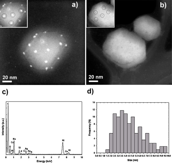
The interaction of nanoparticles with biomolecules and microorganisms is an expanding field of research. Within this field, an area that has been largely unexplored is the interaction of metal nanoparticles with viruses. In this work, we demonstrate that silver nanoparticles undergo a size-dependent interaction with HIV-1, with nanoparticles exclusively in the range of 1-10 nm attached to the virus. The regular spatial arrangement of the attached nanoparticles, the center-to-center distance between nanoparticles, and the fact that the exposed sulfur-bearing residues of the glycoprotein knobs would be attractive sites for nanoparticle interaction suggest that silver nanoparticles interact with the HIV-1 virus via preferential binding to the gp120 glycoprotein knobs. Due to this interaction, silver nanoparticles inhibit the virus from binding to host cells, as demonstrated in vitro.


Figures

Figure 1
Figure 1
Transmission electron microscopy (TEM) of the foamy carbon-coated silver nanoparticles. a) TEM image of the sample prepared by dispersing the as-received powder in deionized water by ultra-sonication. The agglomeration of particles inside the foamy carbon matrix is observed. b) TEM image of nanoparticles outside of the carbon matrix. The broad distribution of shapes can be observed. c)-f) TEM images of nanoparticles with different morphologies. c) Icosahedral. d) Decahedral. e) Elongated. f) Octahedral. g) High Resolution TEM image of the foamy carbon matrix.
Figure 2
Figure 2
Silver nanoparticle preparations. a) TEM image of free surface silver nanoparticles released from the foamy carbon matrix by dispersing the as-received powder in deionized water by ultra-sonication. b) Size distribution of free surface nanoparticles measured by TEM analysis. c) UV-Visible spectrum of carbon-coated silver nanoparticles. d) HAADF image of PVP-coated silver nanoparticles. e) Size distribution of PVP-coated nanoparticles measured by TEM analysis. f) UV-Visible spectrum of PVP-coated silver nanoparticles. g) HAADF image of BSA-coated silver nanoparticles. h) Size distribution of BSA-coated nanoparticles measured by TEM analysis. i) UV-Visible spectrum of BSA-coated silver nanoparticles.
Figure 3
Figure 3
HAADF images of the HIV-1 virus. a) HAADF image of an HIV-1 virus exposed to BSA-conjugated silver nanoparticles. Inset shows the regular spatial arrangement between groups of three nanoparticles. b) HAADF image of HIV-1 viruses without silver nanoparticle treatment. Inset highlight the regular spatial arrangement observed on the surface of the untreated HIV-1 virus. c) EDS analysis of image a) confirming the presence of Ag. The C signal comes from both the TEM grid and the virus, O, and P are from the virus, and Na, Cl, and K are present in the culture medium. Ni and Si come from the TEM grid, while Cu is attributed to the sample holder. d) Composite size distribution of silver nanoparticles bound to the HIV-1 virus, derived from all tested preparations.
Figure 4
Figure 4
Inhibition of HIV-1 and toxicity data. a) Assessment of HIV-1 mediated syncytium formation in MT-2 cells. b) Percentage of HIV-1 transmission in cMAGI cells. The toxicity of the nanoparticle preparations against MT-2 cells was determined using the Trypan Blue exclusion assay. The samples were incubated at 37°C, and the cells were evaluated via optical microscopy after c) 3 h and d) 24 h of exposure to silver nanoparticles.

References

    1. Bonnemann H, Richards RM. Nanoscopic metal particles- Synthetic methods and potential applications. Eur J Inorg Chem. 2001;10:2455–2480. doi: 10.1002/1099-0682(200109)2001:10<2455::AID-EJIC2455>3.3.CO;2-Q. - DOI
    1. Nam JM, Thaxton CS, Mirkin CA. Nanoparticle-based bio-bar codes for the ultrasensitive detection of proteins. Science. 2003;301:1884–1886. doi: 10.1126/science.1088755. - DOI - PubMed
    1. Tkachenko AG, Xie H, Coleman D, Glomm W, Ryan J, Anderson MF, Franzen S, Feldheim DL. Multifunctional Gold Nanoparticle-Peptide Complexes for Nuclear Targeting. J Am Chem Soc. 2003;125:4700–4701. doi: 10.1021/ja0296935. - DOI - PubMed
    1. Hirsch LR, Stafford RJ, Bankson JA, Sershen SR, Rivera B, Price RE, Hazle JD, Halas NJ, West JL. Nanoshell-mediated near-infrared thermal therapy of tumors under magnetic resonance guidance. PNAS. 2003;100:13549–13554. doi: 10.1073/pnas.2232479100. - DOI - PMC - PubMed
    1. Liz-Marzan LM. Nanometals: Formation and color. Materials Today. 2004;7:26–31. doi: 10.1016/S1369-7021(04)00080-X. - DOI

LinkOut - more resources