

Intraoperative and postoperative gamma detection of somatostatin receptors in bone-invasive en plaque meningiomas
- PMID: 15987576
- PMCID: PMC2756774
- DOI: 10.1227/01.neu.0000163490.15578.ff
Intraoperative and postoperative gamma detection of somatostatin receptors in bone-invasive en plaque meningiomas
Abstract
Objective: Scintigraphy with a radiolabeled somatostatin analog ((111)In-diethylenetriaminepenta-acetic acid octreotide) detects the somatostatin receptors that are found in vitro in all meningiomas. Previous studies have proved the benefit of radioimmunoguided surgery, with a hand-held gamma probe, for the assessment and removal of neuroendocrine tumors. We conducted a study to determine whether intraoperative radiodetection of somatostatin receptors is feasible and could increase the probability of complete meningioma resection, especially for bone-invasive en plaque meningiomas, which are difficult to control surgically.
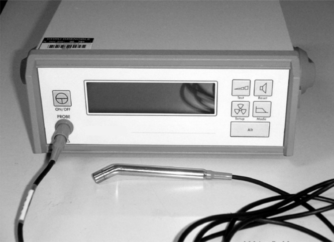
Methods: Eighteen patients with en plaque sphenoid wing and cranial convexity meningiomas were studied by preoperative and postoperative somatostatin receptor scintigraphy. In 10 of them, intraoperative radiodetection with a hand-held gamma probe was performed 24 hours after the intravenous administration of (111)In-diethylenetriaminepenta-acetic acid octreotide. This procedure was combined with a computer-aided navigation system.
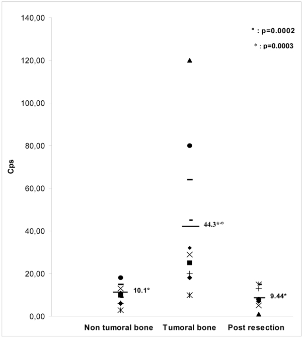
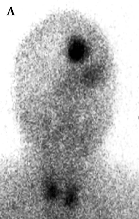
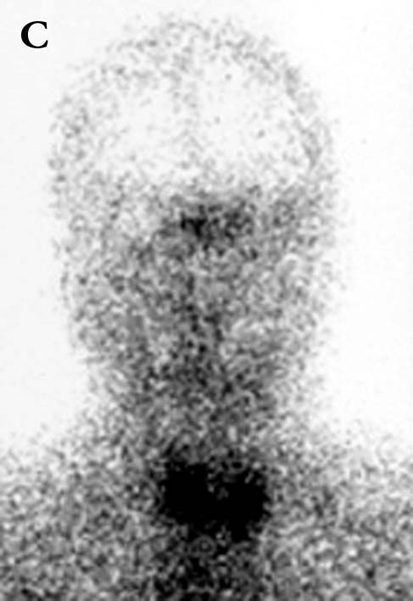
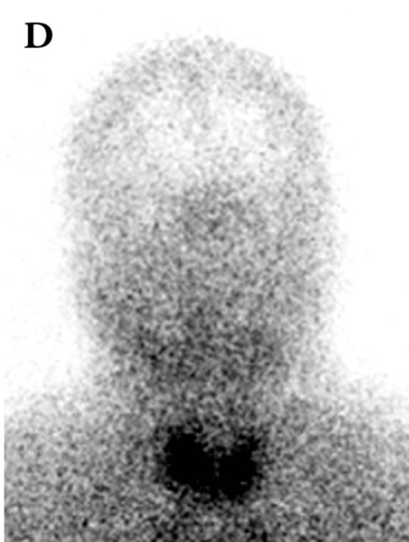
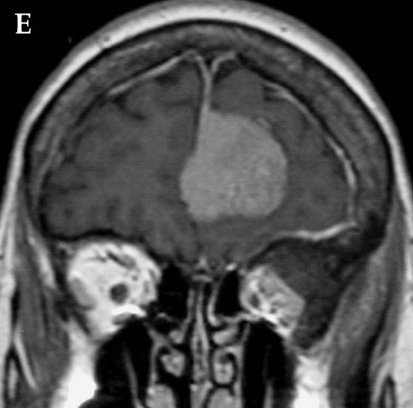
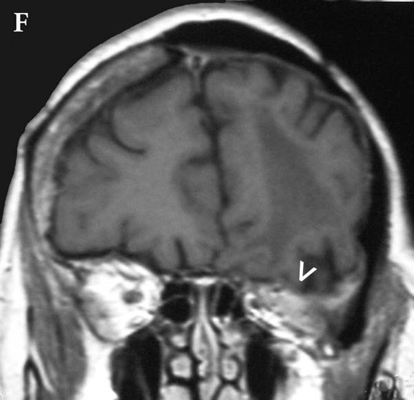
Results: All preoperative scintigrams were positive. Intraoperative gamma probe detection was achieved for the invaded bone, dura, and periorbit of sphenoid wing meningiomas. The average tumor/nontumor count ratio was 2:1, with a maximum of 12:1, thus allowing precise detection capable of defining the tumor margins. In three cases of sphenoid wing meningiomas, postoperative scintigrams were helpful for the determination of recurrences that magnetic resonance imaging failed to detect.
Conclusion: These preliminary data show that intraoperative radiodetection of somatostatin receptors with a hand-held gamma probe is feasible and may be helpful to guide the surgical removal of bone-invasive en plaque meningiomas. Preoperative and postoperative scintigraphy may be useful for the management and follow-up of patients with these tumors.
Figures
Similar articles
-
Radioguided improved resection of a cranial base meningioma.Neurosurgery. 2009 Mar;64(3 Suppl):ons84-5; discussion ons85. doi: 10.1227/01.NEU.0000336309.14805.F5. Neurosurgery. 2009. PMID: 19240580
-
Reduction in the recurrence of meningiomas by combining somatostatin receptor scintigraphy of (99m)Tc-HYNIC-octreotide SPECT/CT and radio guidance with a hand-held γ-probe during surgery.Nucl Med Commun. 2013 Mar;34(3):249-53. doi: 10.1097/MNM.0b013e32835bdfc9. Nucl Med Commun. 2013. PMID: 23276828
-
Intraoperative gamma probe detection of neuroendocrine tumors.J Nucl Med. 1998 Jul;39(7):1155-60. J Nucl Med. 1998. PMID: 9669386
-
Use of radiolabeled somatostatin analogs in the identification and treatment of somatostatin receptor-bearing tumors.Digestion. 1993;54 Suppl 1:88-91. doi: 10.1159/000201084. Digestion. 1993. PMID: 8359574 Review.
-
Radio-guided surgery in neuroendocrine tumors.J Surg Oncol. 2007 Sep 15;96(4):309-15. doi: 10.1002/jso.20868. J Surg Oncol. 2007. PMID: 17726664 Review.
Cited by
-
A comprehensive overview of radioguided surgery using gamma detection probe technology.World J Surg Oncol. 2009 Jan 27;7:11. doi: 10.1186/1477-7819-7-11. World J Surg Oncol. 2009. PMID: 19173715 Free PMC article. Review.
-
Advances in meningioma therapy.Curr Neurol Neurosci Rep. 2009 May;9(3):231-40. doi: 10.1007/s11910-009-0034-5. Curr Neurol Neurosci Rep. 2009. PMID: 19348712 Review.
-
Peptide receptor radionuclide therapy with (90)Y-DOTATOC in recurrent meningioma.Eur J Nucl Med Mol Imaging. 2009 Sep;36(9):1407-16. doi: 10.1007/s00259-009-1115-z. Epub 2009 Mar 25. Eur J Nucl Med Mol Imaging. 2009. PMID: 19319527 Clinical Trial.
-
Theragnostic Use of Radiolabelled Dota-Peptides in Meningioma: From Clinical Demand to Future Applications.Cancers (Basel). 2019 Sep 22;11(10):1412. doi: 10.3390/cancers11101412. Cancers (Basel). 2019. PMID: 31546734 Free PMC article. Review.
-
Management of Recurrent Meningiomas: State of the Art and Perspectives.Cancers (Basel). 2022 Aug 18;14(16):3995. doi: 10.3390/cancers14163995. Cancers (Basel). 2022. PMID: 36010988 Free PMC article. Review.
References
-
- Adams S, Baum RP, Hertel A, Wenisch HJ, Staib-Sebler E, Herrmann G, Encke A, Hor G. Intraoperative gamma probe detection of neuroendocrine tumors. J Nucl Med. 1998;39:1155–1160. - PubMed
-
- Black PM. Meningiomas. Neurosurgery. 1993;32:643–657. - PubMed
-
- Black PM. Hormones, radiosurgery and virtual reality: new aspects of meningioma management. Can J Neurol Sci. 1997;24:302–306. - PubMed
-
- Bohuslavizki KH, Brenner W, Braunsdorf WE, Behnke A, Tinnemeyer S, Hugo HH, Jahn N, Wolf H, Sippel C, Clausen M, Mehdorn HM, Henze E. Somatostatin receptor scintigraphy in the differential diagnosis of meningioma. Nucl Med Commun. 1996;17:302–310. - PubMed
-
- De Menis E, Tulipano G, Villa S, Billeci D, Bonfanti C, Pollara P, Pauletto P, Giustina A. Development of a meningioma in a patient with acromegaly during octreotide treatment: are there any causal relationships? J Endocrinol Invest. 2003;26:359–363. - PubMed
Publication types
MeSH terms
Substances
LinkOut - more resources
Full Text Sources
Research Materials
