. 2005 Sep;16(9):4256-66.
doi: 10.1091/mbc.e05-03-0219. Epub 2005 Jun 29.

Multiple myosin II heavy chain kinases: roles in filament assembly control and proper cytokinesis in Dictyostelium

Affiliations

Multiple myosin II heavy chain kinases: roles in filament assembly control and proper cytokinesis in Dictyostelium

Shigehiko Yumura et al. Mol Biol Cell. 2005 Sep.

Abstract

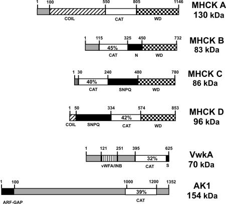
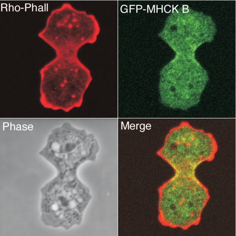
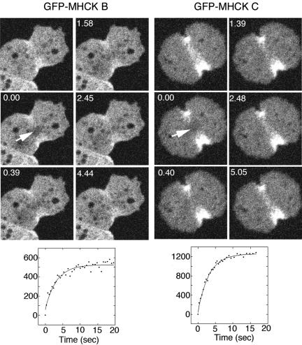
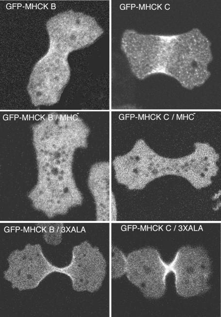
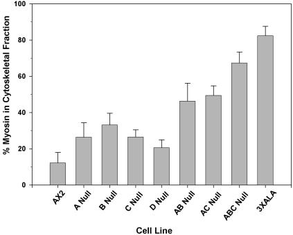
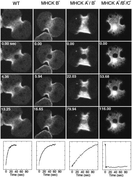
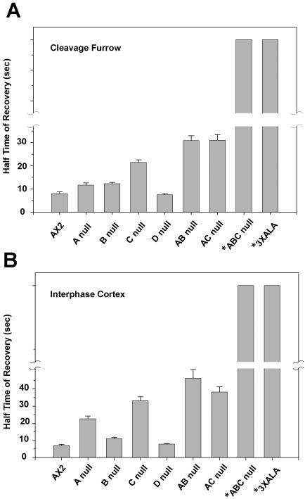
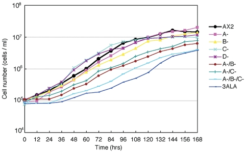
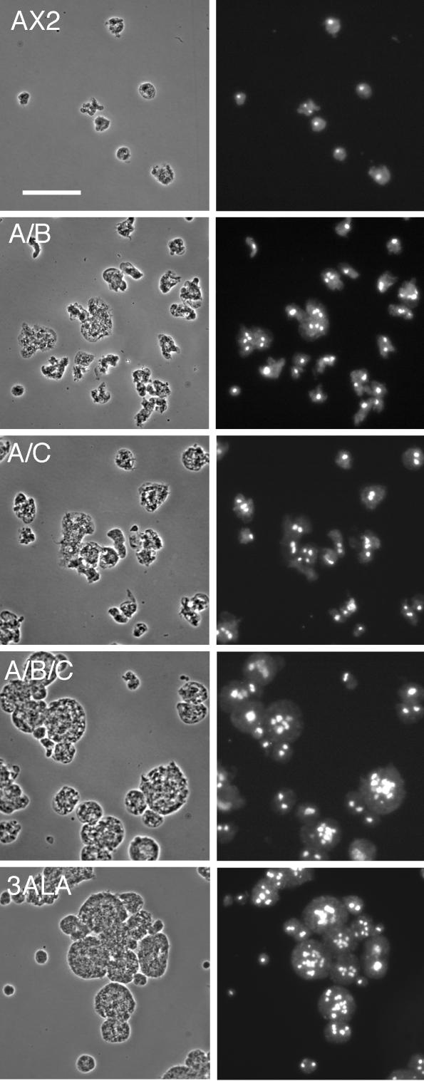
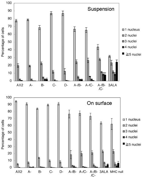
Myosin II filament assembly in Dictyostelium discoideum is regulated via phosphorylation of residues located in the carboxyl-terminal portion of the myosin II heavy chain (MHC) tail. A series of novel protein kinases in this system are capable of phosphorylating these residues in vitro, driving filament disassembly. Previous studies have demonstrated that at least three of these kinases (MHCK A, MHCK B, and MHCK C) display differential localization patterns in living cells. We have created a collection of single, double, and triple gene knockout cell lines for this family of kinases. Analysis of these lines reveals that three MHC kinases appear to represent the majority of cellular activity capable of driving myosin II filament disassembly, and reveals that cytokinesis defects increase with the number of kinases disrupted. Using biochemical fractionation of cytoskeletons and in vivo measurements via fluorescence recovery after photobleaching (FRAP), we find that myosin II overassembly increases incrementally in the mutants, with the MHCK A(-)/B(-)/C(-) triple mutant showing severe myosin II overassembly. These studies suggest that the full complement of MHC kinases that significantly contribute to growth phase and cytokinesis myosin II disassembly in this organism has now been identified.


Figures

Figure 1.
Figure 1.
Domain organization of Dictyostelium complement of alpha kinases. Amino acid residue positions indicated above each sequence. COIL refers to segments with strong predicted or experimentally demonstrated coiled-coil character; CAT, the conserved alpha kinase catalytic domain of each enzyme, with percent identity to MHCK A indicated for each family member; WD, the WD-repeat domain, implicated in targeting MHCKs to the myosin II substrate; N, a polyasparagine-rich segment; SNPQ, a low complexity segment of MHCK C that is rich in serine, asparagines, proline, and glutamine; S, a polyserine segment; vWFA/INB, the vWFA motif/Integrin B motif domain of VwkA.
Figure 2.
Figure 2.
GFP-MHCK B displays enrichment in the contractile ring. Ax2 cells expressing GFP-MHCK B were fixed and permeabilized and then stained with rhodamine-phalloidin.
Figure 3.
Figure 3.
FRAP analysis reveals a high turnover rate for GFP-MHCK B and C associated with the contractile ring. Photobleaching was performed on the contractile ring of an actively dividing cell (0-s time point). Recovery was monitored in this zone >15 s, revealing a half-life of recovery of 1.72 ± 0.30 s (SE; n = 15), and 2.32 ± 0.25 s (SE; n = 18), respectively
Figure 4.
Figure 4.
Distribution of MHCK B and C in the contractile ring depends on myosin II. Cells were fixed and observed by confocal fluorescence microscopy. Neither GFP-MHCK B nor C displayed its localization to the cleavage furrow in myosin heavy chain null cells (MHC-). MHCK B and C still localized at the cleavage furrow in the background of 3XALA myosin II, indicating that these MHCKs recognize a target zone of the myosin II tail that is larger than just the phosphorylation site alone.
Figure 5.
Figure 5.
Assembly of Myosin II into Triton-resistant cytoskeletal fractions. Cells were grown in Petri dishes to near-confluence and then harvested and lysed with Triton X-100. Cytoskeletal ghosts were isolated via centrifugation and SDS-PAGE, and densitometry was used to determine percent of myosin II associated with the cytoskeletal fractions. Bars, SEM; n = 7–9 samples for each cell line.
Figure 6.
Figure 6.
Photobleaching analysis of GFP-myosin II turnover in the contractile ring in single, double, and triple MHCK gene disruption lines. One example of each class of gene disruption is presented. For each series the top image was collected just before photobleaching, and the next three images beneath that present examples of the fluorescence recover time course (time in sec). Below each series is a graph reflecting fluorescence recovery rate for the bleached spot. The X axis is presented in seconds, and the Y axis is arbitrary units reflecting rate of fluorescence recovery. The solid lines were generated by curve-fitting as described in Materials and Methods. These examples illustrate the decrease in GFP-myosin II turnover, which occurs to an increasing degree as more MHCK genes are disrupted. A large spherical fluorescence in the cleavage furrow in the MHCK A-/B-/C- cell was an aggregation of myosin II due to its overassembly. Aggregates of transfected MHC gene constructs have been observed in several previous reports, particularly in the triple MHCK knockout cells and with transfection of 3XALA MHC constructs (Egelhoff et al., 1993; Nagasaki et al., 2002).
Figure 7.
Figure 7.
Graphical comparison of fluorescence recovery times for GFP-myosin II assembled into (A) the contractile ring of dividing cells and (B) the cortex of interphase cells. Error bars, SE of mean. See Tables 1 and 2 for n values. Asterisks indicate that recovery values for these samples could not be determined due to very slow turnover. Bars for these samples indicate only that values are large relative to the other samples.
Figure 8.
Figure 8.
Growth rates in suspension culture for MHCK gene disruption cell lines. Cells collected from Petri dishes were inoculated into shaker flasks at a density of 105 cells/ml and counted daily.
Figure 9.
Figure 9.
Multinucleation in suspension culture of MHCK knockout cell lines. Cells were grown in suspension culture for 3 d, fixed, and stained with DAPI to assess degree of multinucleation. Bar, 50 μm.
Figure 10.
Figure 10.
Multinucleation in MHCK-deficient cells and 3XALA myosin cells is most severe during suspension culture. Cells were collected either from suspension culture as in previous figure or from Petri dishes and processed as in previous figure and then scored for number of nuclei per cell.

References

    1. Abu-Elneel, K., Karchi, M., and Ravid, S. (1996). Dictyostelium myosin II is regulated during chemotaxis by a novel protein kinase C (PKC). J. Biol. Chem. 271, 977-984. - PubMed
    1. Baumann, O. (2004). Spatial pattern of nonmuscle myosin-II distribution during the development of the Drosophila compound eye and implications for retinal morphogenesis. Dev. Biol. 269, 519-533. - PubMed
    1. Betapudi, V., Mason, C., Licate, L., and Egelhoff, T. T. (2005). Identification and Characterization of a novel α-kinase with a von Willebrand factor A-like motif localized to the contractile vacuole and Golgi complex in Dictyostelium discoideum. Mol. Biol. Cell 16, 2248-2262. - PMC - PubMed
    1. Betapudi, V., Shoebotham, K., and Egelhoff, T. T. (2004). Generation of double gene disruptions in Dictyostelium discoideum using a single antibiotic marker selection. Biotechniques 36, 106-112. - PubMed
    1. Bosgraaf, L., Russcher, H., Smith, J. L., Wessels, D., Soll, D. R., and Van Haastert, P. J. (2002). A novel cGMP signalling pathway mediating myosin phosphorylation and chemotaxis in Dictyostelium. EMBO J. 21, 4560-4570. - PMC - PubMed

Publication types

MeSH terms