

doi: 10.1110/ps.051466205.
Epub 2005 Jun 29.
A scalable, GFP-based pipeline for membrane protein overexpression screening and purification
Affiliations
- PMID: 15987891
- PMCID: PMC2279312
- DOI: 10.1110/ps.051466205
Item in Clipboard
A scalable, GFP-based pipeline for membrane protein overexpression screening and purification
Protein Sci.
2005 Aug.
Abstract
We describe a generic, GFP-based pipeline for membrane protein overexpression and purification in Escherichia coli. We exemplify the use of the pipeline by the identification and characterization of E. coli YedZ, a new, membrane-integral flavocytochrome. The approach is scalable and suitable for high-throughput applications. The GFP-based pipeline will facilitate the characterization of the E. coli membrane proteome and serves as an important reference for the characterization of other membrane proteomes.
Figures
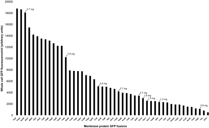
GFP-based MP overexpression screen. Forty-eight E. coli MPs were overexpressed as GFP fusions in E. coli as described in Materials and Methods. Whole-cell GFP fluorescence is expressed in arbitrary units. Nine MP-GFP fusions were isolated by using a standard protocol as described in Materials and Methods. The amount of purified MP-GFP fusion that can be isolated from 1 L of culture is shown in milligrams.

Digestion of MP-GFP fusions and characterization of recovered proteins. (A) SDS-PAGE of purified and TEV-digested YbaT- (left), GltP- (middle), and YedZ- (right) GFP fusions as described in Materials and Methods: purified fusion (lane 1), TEV protease (lane 2), eluted batch bound material (lane 3; GFP is marked with *), and recovered membrane protein (lane 4). The gel was stained with Coomassie brilliant blue R-250. (B) Cleavage of YbaT-GFP with TEV protease monitored under UV light: YbaT-GFP (lane 1), batch bound material (lane 2), and recovered YbaT (lane 3). (C) Glutamate uptake activity of proteoliposomes containing purified GltP recovered from a GltP-GFP fusion (diamonds), purified GltP-His8 (squares), and control liposomes containing no protein (triangles). (D) Purified YedZ under ambient light.

Optical absorption spectra of YedZ and identification of its cofactors. (A) Optical spectra of purified YedZ (concentration of ~0.01 mg/ml) after oxidation with ferricyanide and reduction with sodium dithionite. The absorption maxima of the reduced form of YedZ are α=558 nm, β=528 nm, and γ=425 nm. (B) MALDI-TOF MS spectrum of purified YedZ in reflectron mode. Measurements were in them/z range from 500–1000mass units. The mass of 617,47 represents the singly charged ion of heme b. (C) Identification by reverse-phase liquid chromatography of the flavin FMN in the supernatant from TCA-precipitated YedZ.

Functional overexpression of the human KDEL-receptor as a GFP fusion in L. lactis. (A) Western blot probed with antibodies raised against peptide 192–212 of the KDEL-receptor: membranes of L. lactis cells overexpressing the KDEL-receptor (lane 1) and membranes of L. lactis cells overexpressing a KDEL-GFP fusion (lane 2). (B) KDELr ligand binding studies carried out with isolated membranes of the uninduced (lane 1) and induced (lane 2) control strain (harboring an empty expression vector), the uninduced (lane 3) and induced (lane 4) strain with a KDELr expression vector (Kunji et al. 2003), and the uninduced (lane 5) and induced (lane 6) strain with the KDELr-GFP fusion expression vector. Binding assays were carried out in duplicate.
References
-
- Barber, M.J., Desai, S.K., Marohnic, C.C., Hernandez, H.H., and Pollock, V.V. 2002. Synthesis and bacterial expression of a gene encoding the heme domain of assimilatory nitrate reductase. Arch. Biochem. Biophys. 402 38–50. - PubMed
-
- Burch, H.B. 1957. Fluorometric assay of FAD, FMN, and riboflavin. Methods Enzymol. 3 960–962.
-
- Daley, D.O., Rapp, M., Granseth, E., Melén, K., Drew, D., and von Heijne, G. 2005. Global topology analysis of the Escherichia coli inner membrane proteome. Science 308 1321–1323. - PubMed
-
- Drew, D.E., von Heijne, G., Nordlund, P., and de Gier, J.W. 2001. Green fluorescent protein as an indicator to monitor membrane protein overexpression in Escherichia coli. FEBS Lett. 507 220–224. - PubMed
Publication types
MeSH terms
Substances
Grants and funding
LinkOut - more resources
Full Text Sources
Other Literature Sources
Molecular Biology Databases
