Review
. 2005 Jul;14(7):1697-709.
doi: 10.1110/ps.051406805.

Not all J domains are created equal: implications for the specificity of Hsp40-Hsp70 interactions

Affiliations
Review

Not all J domains are created equal: implications for the specificity of Hsp40-Hsp70 interactions

Fritha Hennessy et al. Protein Sci. 2005 Jul.

Abstract

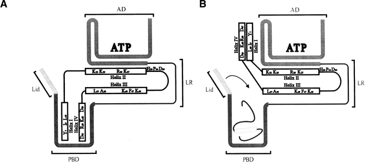
Heat shock protein 40s (Hsp40s) and heat shock protein 70s (Hsp70s) form chaperone partnerships that are key components of cellular chaperone networks involved in facilitating the correct folding of a broad range of client proteins. While the Hsp40 family of proteins is highly diverse with multiple forms occurring in any particular cell or compartment, all its members are characterized by a J domain that directs their interaction with a partner Hsp70. Specific Hsp40-Hsp70 chaperone partnerships have been identified that are dedicated to the correct folding of distinct subsets of client proteins. The elucidation of the mechanism by which these specific Hsp40-Hsp70 partnerships are formed will greatly enhance our understanding of the way in which chaperone pathways are integrated into finely regulated protein folding networks. From in silico analyses, domain swapping and rational protein engineering experiments, evidence has accumulated that indicates that J domains contain key specificity determinants. This review will critically discuss the current understanding of the structural features of J domains that determine the specificity of interaction between Hsp40 proteins and their partner Hsp70s. We also propose a model in which the J domain is able to integrate specificity and chaperone activity.


Figures

Figure 1.
Figure 1.
Structural motifs found in E. coli DnaJ. (A) A diagrammatic representation of the domains present in E. coli DnaJ. J represents the J domain, G/F stands for the Gly/Phe-rich region, and cysteine repeats represents the four zinc-finger-like motifs. The first three domains correspond to approximately half DnaJ. (B) A ribbon representation of the J domain (1XBL) (Pellechia et al. 1996). The conserved HPD motif is depicted in purple. The four helices are labeled. (C) A ribbon representation of the cysteine repeats (1EXK) (Martinez-Yamout et al. 2000). Cysteine residues are depicted in red (repeats 1 and 2) and blue (repeats 3 and 4). Repeats 1 and 4 form zinc center 1 and repeats 2 and 3 form zinc center 2 (Linke et al. 2003). The coordinated zinc atoms are shown in CPK format. This diagram is not to scale. The figures were generated using Molscript (Kraulis 1991).
Figure 2.
Figure 2.
Ribbon representation of the structures of J domains from various Hsp40 and Hsp40-like proteins. The structures for E. coli DnaJ (1XBL, 1BQZ, 1BQ0) (Pellecchia et al. 1996; Huang et al. 1998), human HDJ1 (1HDJ) (Qian et al. 1996), and the murine polyomavirus T antigen (1FAF) (Berjanskii et al. 2000) J domains were determined using NMR. The structure of E. coli Hsc20 (1FPO) (Cupp-Vickery and Vickery 2000), SV40 T antigen (1GH6) (Kim et al. 2001), and bovine auxilin (1NZ6) (Jiang et al. 2003) J domains were determined using X-ray crystallography. The structures were visualized using Molscript (Kraulis 1991). The HPD motif is depicted in purple. Helices II and III are in blue, with the more mobile helices I and IV in red. The helices and HPD motif are labeled on the DnaJ 1XBL J domain structure. Only the J domain region is shown even if additional structural regions were determined.
Figure 3.
Figure 3.
Schematic showing the protein folding cycle involving the interaction of Hsp40 and Hsp40-like proteins with partner Hsp70s. Pi is inorganic phosphate. Dotted lines indicate the two different paths by which client proteins and Hsp40 or Hsp40-like proteins can enter the cycle. Client proteins are either recognized by Hsp70, following which an Hsp40 or Hsp40-like protein enters the cycle, or are presented to Hsp70 by an Hsp40 or Hsp40-like protein. ATP hydrolysis, stimulated by an Hsp40 or Hsp40-like protein, causes a conformational shift in the peptide binding domain, locking in the client protein. Nucleotide exchange then reverses the conformational shift, allowing for the release and subsequent folding of the client protein. Alternatively the client protein can re-enter the cycle.
Figure 4.
Figure 4.
Specificity of interaction between Hsp70 and Hsp40 or Hsp40-like proteins in E. coli. Thick arrows indicate known partnerships. Dotted lines and arrows indicate lower levels of ATP hydrolysis stimulation in vitro. Thin lines with squares replacing arrowheads indicate no detectable levels of ATPase stimulation and no known partnership. “Hsp40/Hsp40-like” indicates the list of different E. coli Hsp40 or Hsp40-like proteins; “Hsp70” indicates the list of different E. coli Hsp70 proteins.
Figure 5.
Figure 5.
Model for the interaction of the J domain with Hsp70. Residues important in J domain–based interactions with Hsp70 are shown using the single letter code. The J domain has four helices labeled I–IV. Charged residues in helices II and III interact with the underside of the Hsp70 ATPase domain (AD). (A) helices I and IV possibly act as a client protein mimic and interact transiently with the Hsp70 peptide binding domain (PBD). (B) The correct client protein at the correct concentration is able to displace helices I and IV and bind in the PBD; this interaction is stabilized by subsequent ATP hydrolysis and PBD lid closure (indicated by the downward arrow). The Hsp70 linker region is designated LR.

References

    1. Auger, I. and Roudier, J. 1997. A function for the QKRAA amino acid motif: Mediating binding of DnaJ to DnaK. Implications for the association of rheumatoid arthritis with HLA-DR4. J. Clin. Invest. 99 1818–1822. - PMC - PubMed
    1. Berjanskii, M.V., Riley, M.I., Xie, A., Semenchenko, V., Folk, W.R., and Van Doren, S.R. 2000. NMR structure of the N-terminal J domain of murine polyomavirus T antigens. J. Biol. Chem. 275 36094–36103. - PubMed
    1. Berjanskii, M.V., Riley, M.I., and Van Doren, S.R. 2002. Hsc70-interacting HPD loop of the J domain of polyomavirus T antigens fluctuates in ps to ns and μs to ms. J. Mol. Biol. 321 503–516. - PubMed
    1. Bolliger, L., Deloche, O., Glick, B.S., Georgopoulos, C., Jenö, P., Kronidou, N., Horst, M., Morishima, N., and Schatz, G. 1994. A mitochondrial homologue of bacterial GrpE interacts with mitochondrial hsp70 and is essential for viability. EMBO J. 13 1998–2006. - PMC - PubMed
    1. Bork, P., Sander, C., Valencia, A., and Bukau, B. 1992. A module of the DnaJ heat shock proteins found in malaria parasites. Trends Biochem. Sci. 17 129. - PubMed

Publication types

MeSH terms

LinkOut - more resources