. 2005 Jun 29;25(26):6251-9.
doi: 10.1523/JNEUROSCI.4601-04.2005.

Brain-derived neurotrophic factor is required for the establishment of the proper number of dopaminergic neurons in the substantia nigra pars compacta

Affiliations

Brain-derived neurotrophic factor is required for the establishment of the proper number of dopaminergic neurons in the substantia nigra pars compacta

Zachary C Baquet et al. J Neurosci. .

Abstract

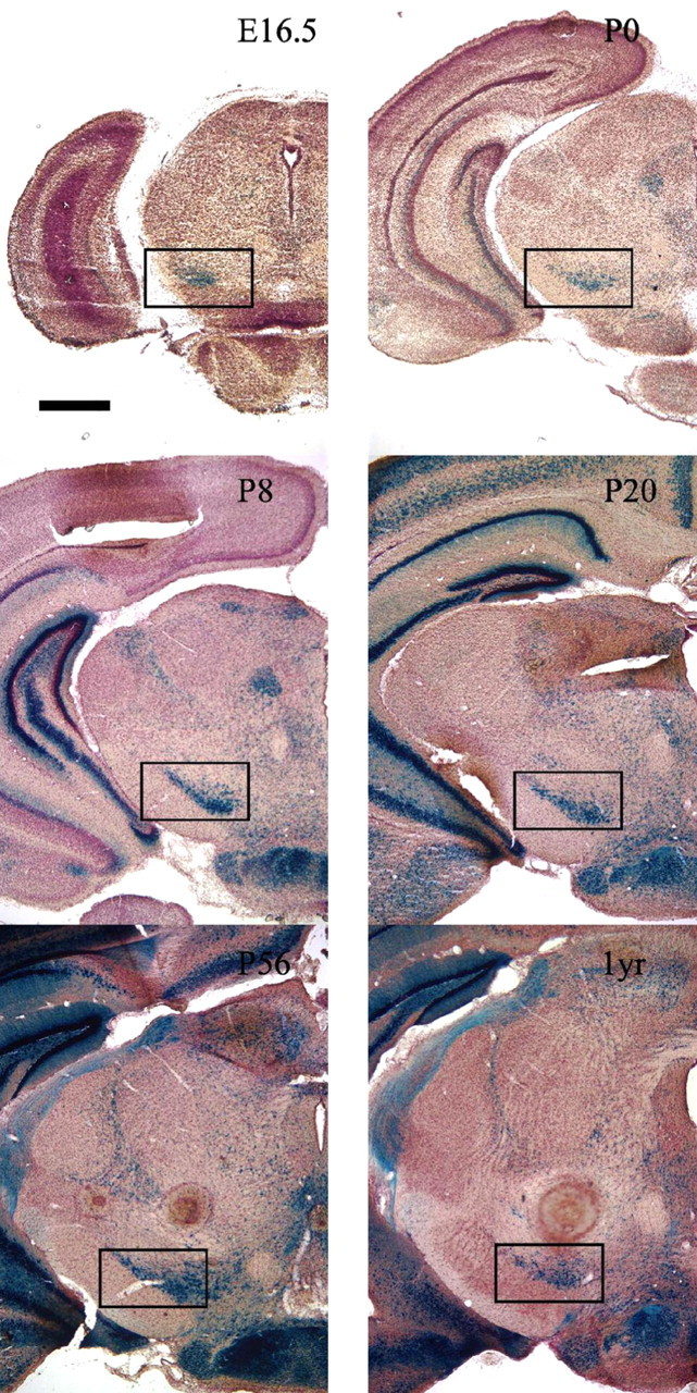
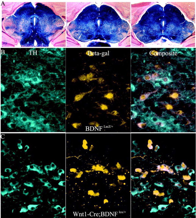
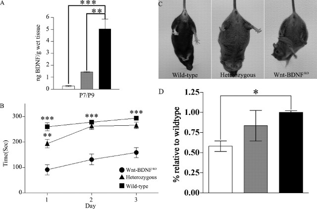
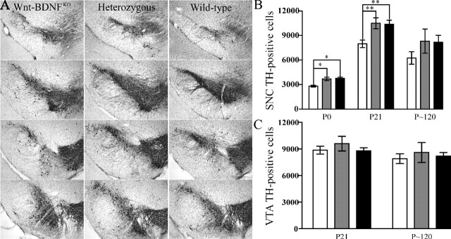
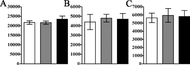
Brain-derived neurotrophic factor (BDNF) has been implicated in regulating neuronal survival, differentiation, and synaptic plasticity. Reduced expression of BDNF within the substantia nigra accompanies the deterioration of dopaminergic neurons in Parkinson's disease (PD) patients. Analysis of the effects of long-term BDNF absence from the CNS has been difficult because of the early postnatal lethality of BDNF-/- mice. Mice with a floxed BDNF allele were bred with Wnt1-Cre mice to generate Wnt-BDNF(KO) mice that lack BDNF from the midbrain-hindbrain (MHB). These mice are viable but exhibit hindlimb clutching and poor rotarod performance. Tyrosine hydroxylase (TH)-positive neuron numbers in the substantia nigra pars compacta (SNC) were estimated using stereological methods, revealing a persistent approximately 23% reduction of these cells at postnatal day 21 (P21) in Wnt-BDNF(KO) mice compared with controls. The diminishment of TH-expressing neurons was present at birth and continued through P120. This deficit appears selective for the dopaminergic population, because at P21, total neuron number within the SNC, defined as neuronal nuclei protein-positive cells, was not significantly reduced. Interestingly, and similar to observations in PD patients, SNC neuron subpopulations are not equally affected. Calbindin- and calretinin-expressing SNC populations show no significant difference between Wnt-BDNF(KO) mice and controls. Thus, BDNF depletion from the MHB selectively leads to reduced TH expression in a subpopulation of neurons, but it remains unclear whether these cells are lost.


Figures

Figure 1.
Figure 1.
BDNFLacZ expression in the substantia nigra. BDNFLacZ/+ embryos, neonates, and postnatal mice demonstrate the presence of BDNF within the substantia nigra pars compacta. The black box indicates the location of the SNC. Coronal sections (40 μm) of different ages, starting at E16.5 and ending at 1 year (1yr) (indicated in the top right portion of each panel), were stained with X-gal, counterstained with Neutral Red, and then photographed at 20× magnification. Scale bar, 1 mm.
Figure 2.
Figure 2.
Expression of Cre in midbrain BDNF-expressing neurons. A, X-gal stain of 40 μm coronal sections (counterstained with Neutral Red) from the midbrain-hindbrain of P21 Wnt1-Cre;R26R mice. The dark blue stain indicates areas of Cre activity. Outside the MHB, there are only scattered CNS cells that undergo recombination. B, Immunofluorescent double labeling using antibodies to tyrosine hydroxylase and β-gal of 10 μm cryostat sections from the anterior portion of the SNC of a BDNFLacZ/+ mouse documents expression in multiple SNC cells. Images were photographed at 400× magnification. Lateral is to the left of the image, and the midline is to the right of the image. C, Effectiveness of Wnt1-Cre at excising BDNF from DA neurons. Images of sections are processed as in B of the SNC of a Wnt1-Cre; BDNFlox/+ mouse.
Figure 3.
Figure 3.
Reduced BDNF protein in MHB leads to motor deficits and reduced striatal TH in Wnt-BDNFKO mice. A, BDNF protein was quantified by ELISA for MHB and expressed as nanograms of BDNF protein per gram of wet tissue. These extracts were obtained from P7 and P9 mice and pooled to increase the n (for P7/P9, n = 5 for Wnt-BDNFKO and wild-type; n = 4 for heterozygous; **p < 0.01; ***p < 0.001; one-way ANOVA with a Newman-Keuls post hoc test). B, Performance of 4- to 5-week-old mice on an accelerating rotarod. Each mouse had three trials each day, which were averaged for the day (Wnt-BDNFKO, n = 7; heterozygous, n = 16; wild type, n = 6; **p < 0.01 heterozygous vs wild type for day 1 only; ***p < 0.001 Wnt-BDNFKO vs heterozygous and wild type all 3 d). C, Wnt-BDNFKO, heterozygous, and wild-type mice at 1, 2, and 4 months of age were suspended by their tails for 1 min. Clasping was defined as the balling up of one or both of the hindlimb paws, accompanied by pulling them into the body and movement of the limbs toward the midline. Four-month-old wild-type, heterozygous, and Wnt-BDNFKO mice undergoing tail suspension are shown. D, Western blot analysis of TH levels in total striatal protein from P35 Wnt-BDNFKO and control mice (n = 4 for Wnt-BDNFKO and wild type; n = 3 for heterozygous; *p < 0.05; one-way ANOVA with a Newman-Keuls post hoc test). White bar, Wnt-BDNFKO; gray bar, heterozygous; black bar, wild type.
Figure 4.
Figure 4.
Wnt-BDNFKO mice have reduced TH expression in the SNC but not in the VTA. A, The extent of the SNC appears reduced in P21Wnt-BDNFKO mice compared with controls, and the number of TH fibers also seems to be reduced. The most anterior section is at the top, and the most posterior section is at the bottom. Coronal cryostat sections (40 μm) taken at 240 μm intervals stained for TH and counterstained with cresyl violet are shown. B, Optical fractionator estimates of the number of DA neurons in both hemispheres of the SNC of Wnt-BDNFKO mice and controls at P0, P21, and P120 (P0, n = 3/genotype; P21, n = 9, 8, and 9 for Wnt-BDNFKO, heterozygous, and wild type, respectively; P120, n = 3/genotype; *p < 0.05; **p < 0.01; one-way ANOVA with a Newman-Keuls posthoc test). C, There is no significant reduction in the total number of TH-positive cells of both hemispheres of the VTA of Wnt-BDNFKO mice compared with controls (P21, n = 11, 7, and 8 for Wnt-BDNFKO, heterozygous, and wild type, respectively, p = 0.6454; P120, n = 3/genotype, p = 0.8170). White bars, Wnt-BDNFKO; gray bars, heterozygous; black bars, wild type.
Figure 5.
Figure 5.
NeuN-positive neuron loss in the SNC of Wnt-BDNFKO mice was not detected. A, NeuN-positive cells were counted in the SNC, defined by outlining the TH-stained population of neurons on adjacent sections, and estimates were obtained for total numbers of neurons in the SNC at P21 (n = 4 for Wnt-BDNFKO and heterozygous; n = 3 for wild type; p = 0.5093). B, Stereological estimates of calbindin-positive cell population in the SNC. C, Estimates of calretinin-positive cell population in the SNC. For both experiments, n = 3/genotype. No significant difference was found between genotypes by one-way ANOVA. White bars, Wnt-BDNFKO; gray bars, heterozygous; black bars, wild type.

References

    1. Auerbach W, Hurlbert MS, Hilditch-Maguire P, Wadghiri YZ, Wheeler VC, Cohen SI, Joyner AL, MacDonald ME, Turnbull DH (2001) The HD mutation causes progressive lethal neurological disease in mice expressing reduced levels of huntingtin. Hum Mol Genet 10: 2515-2523. - PubMed
    1. Baker SA, Stanford LE, Brown RE, Hagg T (2005) Maturation but not survival of dopaminergic nigrostriatal neurons is affected in developing and aging BDNF-deficient mice. Brain Res 1039: 177-188. - PubMed
    1. Baquet ZC, Gorski JA, Jones KR (2004) Early striatal dendrite deficits followed by neuron loss with advanced age in the absence of anterograde cortical brain-derived neurotrophic factor. J Neurosci 24: 4250-4258. - PMC - PubMed
    1. Bayer SA, Wills KV, Triarhou LC, Ghetti B (1995) Time of neuron origin and gradients of neurogenesis in midbrain dopaminergic neurons in the mouse. Exp Brain Res 105: 191-199. - PubMed
    1. Bennett JL, Zeiler SR, Jones KR (1999) Patterned expression of BDNF and NT-3 in the retina and anterior segment of the developing mammalian eye. Invest Ophthalmol Vis Sci 40: 2996-3005. - PubMed

Publication types

MeSH terms