. 2005 Jul;25(14):5973-84.
doi: 10.1128/MCB.25.14.5973-5984.2005.

Aberrant morphology and residual transmitter release at the Munc13-deficient mouse neuromuscular synapse

Affiliations

Aberrant morphology and residual transmitter release at the Munc13-deficient mouse neuromuscular synapse

Frédérique Varoqueaux et al. Mol Cell Biol. 2005 Jul.

Abstract

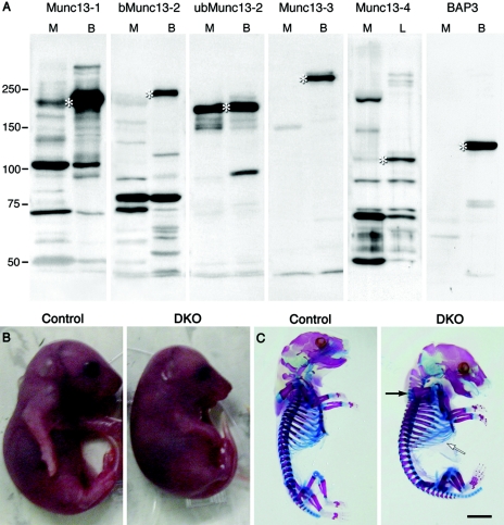
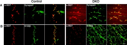
In cultured hippocampal neurons, synaptogenesis is largely independent of synaptic transmission, while several accounts in the literature indicate that synaptogenesis at cholinergic neuromuscular junctions in mammals appears to partially depend on synaptic activity. To systematically examine the role of synaptic activity in synaptogenesis at the neuromuscular junction, we investigated neuromuscular synaptogenesis and neurotransmitter release of mice lacking all synaptic vesicle priming proteins of the Munc13 family. Munc13-deficient mice are completely paralyzed at birth and die immediately, but form specialized neuromuscular endplates that display typical synaptic features. However, the distribution, number, size, and shape of these synapses, as well as the number of motor neurons they originate from and the maturation state of muscle cells, are profoundly altered. Surprisingly, Munc13-deficient synapses exhibit significantly increased spontaneous quantal acetylcholine release, although fewer fusion-competent synaptic vesicles are present and nerve stimulation-evoked secretion is hardly elicitable and strongly reduced in magnitude. We conclude that the residual transmitter release in Munc13-deficient mice is not sufficient to sustain normal synaptogenesis at the neuromuscular junction, essentially causing morphological aberrations that are also seen upon total blockade of neuromuscular transmission in other genetic models. Our data confirm the importance of Munc13 proteins in synaptic vesicle priming at the neuromuscular junction but indicate also that priming at this synapse may differ from priming at glutamatergic and gamma-aminobutyric acid-ergic synapses and is partly Munc13 independent. Thus, non-Munc13 priming proteins exist at this synapse or vesicle priming occurs in part spontaneously: i.e., without dedicated priming proteins in the release machinery.


Figures

FIG. 1.
FIG. 1.
Munc13 isoforms at the neuromuscular junction and phenotypic alterations in the Munc13-1/2-DKO mouse mutant. (A) Immunoblot analysis of muscle membrane extract (M) with anti-Munc13-1, -b/ubMunc13-2, -Munc13-3, -Munc13-4 and -BAP3 isoform-specific antibodies. Brain (lanes B) or lung (lanes L) homogenates were used as positive control. (B and C) E18.5 Munc13-1/2-DKO mutant and control littermate mice gross morphology (B) and skeleton (C; bones are stained in blue and cartilage in pink). A white arrow points to a broadened rib cage, and a black arrow points to a stiffened neck and a compacted spinal cord. Scale bar, 3 mm.
FIG. 2.
FIG. 2.
Impaired morphology of the Munc13-1/2-DKO diaphragm muscle. (A and B) Detail of whole mount (A) and Nissl-stained semithin cross-section (B) of Munc13-1/2-DKO mutant and control littermate. Black arrows indicate oversized blood vessels, and white arrows indicate poorly differentiated myotubes. Scale bars, 100 μm in panel A and 30 μm in panel B.
FIG. 3.
FIG. 3.
Impaired branching and endplate distribution of the Munc13-1/2-DKO phrenic nerve terminating onto the diaphragm surface. (A and B) Detail of the left (A) and right (B) hemidiaphragms of Munc13-1/2-DKO mutant and control littermate, stained for acetylcholinesterase. Scale bar, 180 μm.
FIG. 4.
FIG. 4.
Normal apposition of presynaptic, postsynaptic, and glial elements at the Munc13-1/2-DKO motor endplate. (A and B) Confocal micrographs of Munc13-1/2-DKO mutant and control littermate, double immunostained for α-bungarotoxin (α-BGT; to visualize acetylcholine receptors) and synapsin (as a marker for presynapses) (A) or S-100 (S100β; as a marker for Schwann cells) (B). Scale bars, 90 μm in panel A and 190 μm in panel B.
FIG.5.
FIG.5.
Increased number of motor neurons in the Munc13-1/2-DKO mutant spinal cord. (A) Groups of large-bodied motor neurons are easily identified in the ventral horn of Nissl-stained cervical spinal cord sections of Munc13-1/2-DKO mutant and control littermate. (B) Detail of the motor neuron-specific cholinergic innervation obtained by immunostaining for vAChT in the ventral horn of the spinal cord of Munc13-1/2-DKO mutant and control littermate. (C) Low-magnification electron micrographs of transversally cut right phrenic nerves of Munc13-1/2-DKO mutant and control littermate; the insert shows a detail of a myelinated axon. (D) Quantification of the number of motor neuron axons in the phrenic nerve of control littermate (n = 8) and Munc13-1/2-DKO mutant embryos (n = 6). Scale bars: 170 μm in panel A, 60 μm in panel B, and 7 μm in panel C.
FIG.6.
FIG.6.
Strongly reduced acetylcholine release evoked by nerve impulses, α-latrotoxin, or hypertonic medium at Munc13-1/2-DKO NMJs. Bar graphs display the group mean values ± standard error of the mean (n = 5 to 15 embryos, 1 to 21 NMJs sampled per muscle). (A) Spontaneous uniquantal acetylcholine release, MEPPs, recorded in normal Ringer's medium. Superimposed example traces show the MEPPs observed during a 135-s measuring period. (B) MEPPs recorded in the presence of 2.5 nM α-latrotoxin. (C) MEPPs recorded in the presence of 0.5 M sucrose-Ringer's medium. (D) Examples of nerve stimulation-evoked responses. The moment of phrenic nerve stimulation is indicated with a black triangle. At relative hyperpolarized membrane potentials, a full-size muscle action potential is elicited in control muscle (upper left), leading to contraction that is visible as an artifact on the signal (indicated by open triangle). At Munc13-1/2-DKO NMJs, subthreshold and delayed EPPs and failures were observed (upper right), sometimes leading to delayed muscle action potentials. Subsequent traces (0.3-Hz stimulation) have been superimposed. At depolarized muscle fibers, EPPs become unmasked. At control NMJs, no failures were observed at 0.3-Hz stimulation (bottom left), while at Munc13-1/2-DKO NMJs there were many failures and very small, delayed EPPs. (E) Percentage of stimuli leading to failures. (F) EPP amplitude, normalized to −75 mV membrane potential, failures taken into account. (G) Quantal content (i.e., the number of acetylcholine quanta released upon a single nerve impulse).
FIG. 7.
FIG. 7.
Immature but well-formed neuromuscular synapses in the Munc13-1/2-DKO mutant. Electron micrographs of representative motor endplates in Munc13-1/2-DKO mutant and control littermate. At low magnification (A), Munc13-1/2-DKO motor endplates are always composed of more presynaptic elements, containing numerous small synaptic vesicles, than the littermate ones. In either case, magnified areas of the synaptic active zone (B) allow recognition of small synaptic vesicles docked at the active zone membrane, large dense-core vesicles, clathrin-coated vesicles, and an intact basal lamina. However, the postsynaptic membrane of the Munc13-1/2-DKO muscle cell fails to develop secondary folds that normally accompany the maturation process of neuromuscular synapses (arrow in control). Scale bars: 700 nm in upper panels and 220 nm in lower panels.

References

    1. Allan, D. W., and J. J. Greer. 1997. Development of phrenic motoneuron morphology in the fetal rat. J. Comp. Neurol. 382:469-479. - PubMed
    1. Aravamudan, B., T. Fergestad, W. S. Davis, C. K. Rodesch, and K. Broadie. 1999. Drosophila UNC-13 is essential for synaptic transmission. Nat. Neurosci. 2:965-971. - PubMed
    1. Ashery, U., F. Varoqueaux, T. Voets, A. Betz, P. Thakur, H. Koch, E. Neher, N. Brose, and J. Rettig. 2000. Munc13-1 acts as a priming factor for large dense-core vesicles in bovine chromaffin cells. EMBO J. 19:3586-3596. - PMC - PubMed
    1. Augustin, I., A. Betz, C. Herrmann, T. Jo, and N. Brose. 1999. Differential expression of two novel Munc13 proteins in rat brain. Biochem. J. 337:363-371. - PMC - PubMed
    1. Augustin, I., S. Korte, M. Rickmann, H. A. Kretzschmar, T. C. Sudhof, J. W. Herms, and N. Brose. 2001. The cerebellum-specific Munc13 isoform Munc13-3 regulates cerebellar synaptic transmission and motor learning in mice. J. Neurosci. 21:10-17. - PMC - PubMed

Publication types

MeSH terms