. 2005 Jul;25(14):6047-64.
doi: 10.1128/MCB.25.14.6047-6064.2005.

Activation of a nuclear Cdc2-related kinase within a mitogen-activated protein kinase-like TDY motif by autophosphorylation and cyclin-dependent protein kinase-activating kinase

Affiliations

Activation of a nuclear Cdc2-related kinase within a mitogen-activated protein kinase-like TDY motif by autophosphorylation and cyclin-dependent protein kinase-activating kinase

Zheng Fu et al. Mol Cell Biol. 2005 Jul.

Abstract

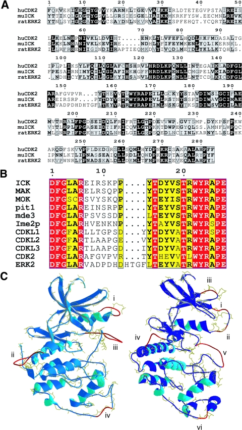
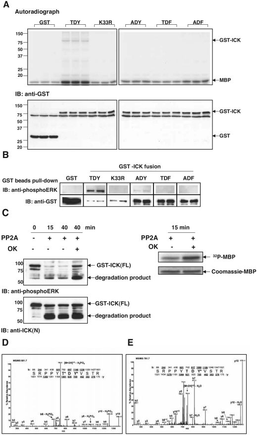
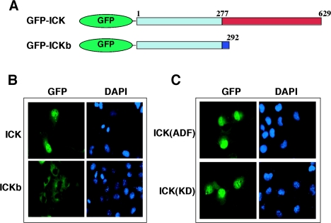
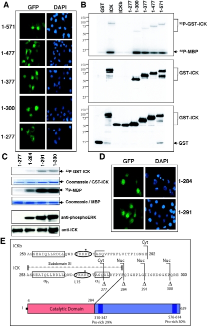
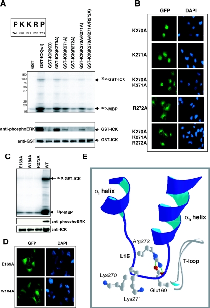
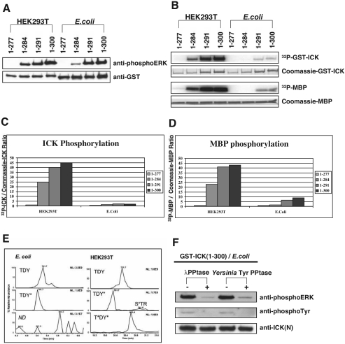
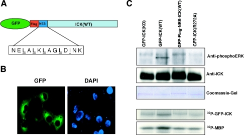
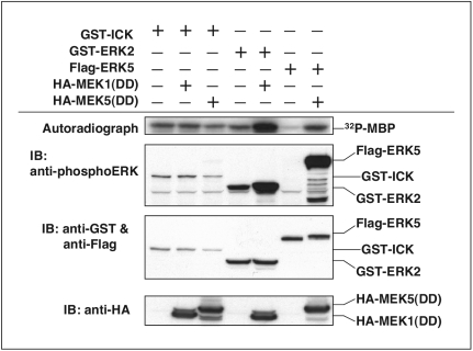
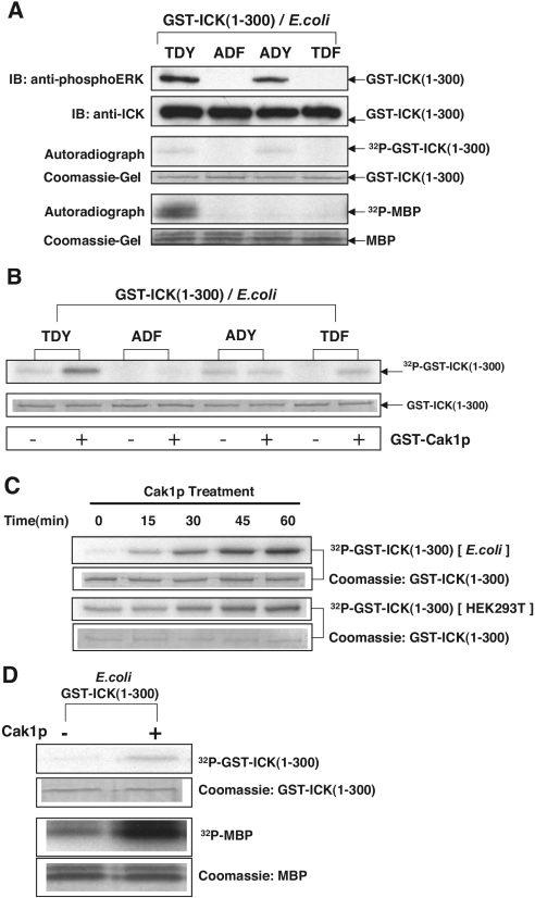
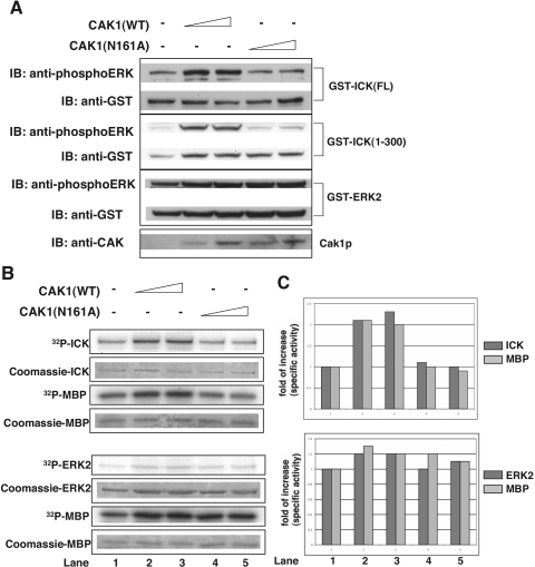
Male germ cell-associated kinase (MAK) and intestinal cell kinase (ICK) are nuclear Cdc2-related kinases with nearly identical N-terminal catalytic domains and more divergent C-terminal noncatalytic domains. The catalytic domain is also related to mitogen-activated protein kinases (MAPKs) and contains a corresponding TDY motif. Nuclear localization of ICK requires subdomain XI and interactions of the conserved Arg-272, but not kinase activity or, surprisingly, any of the noncatalytic domain. Further, nuclear localization of ICK is required for its activation. ICK is activated by dual phosphorylation of the TDY motif. Phosphorylation of Tyr-159 in the TDY motif requires ICK autokinase activity but confers only basal kinase activity. Full activation requires additional phosphorylation of Thr-157 in the TDY motif. Coexpression of ICK with constitutively active MEK1 or MEK5 fails to increase ICK phosphorylation or activity, suggesting that MEKs are not involved. ICK and MAK are related to Ime2p in budding yeast, and cyclin-dependent protein kinase-activating kinase Cak1p has been placed genetically upstream of Ime2p. Recombinant Cak1p phosphorylates Thr-157 in the TDY motif of recombinant ICK and activates its activity in vitro. Coexpression of ICK with wild-type CAK1 but not kinase-inactive CAK1 in cells also increases ICK phosphorylation and activity. Our studies establish ICK as the prototype for a new group of MAPK-like kinases requiring dual phosphorylation at TDY motifs.


Figures

FIG.1.
FIG.1.
ICK is a MAPK- and CDK-like kinase with a TDY motif. (A) Sequence alignment of the catalytic domains of mouse ICK, rat ERK2, and human CDK2 by using Clustal. ICK shares about 38% to 40% overall identity to ERK2 and CDK2 in the catalytic domains shown. ICK, MAK, and MOK have short N termini like CDK2, each only three residues (not shown). ICK also contains a C-terminal domain (not shown) that is similar to that of MAK but more divergent. Numbering corresponds to the catalytic domain residues. (B) Alignment of the T loops of TDY motif kinases in the human genome. Ime2p, pit1, and mde3 are putative homologs of ICK and MAK in yeast. (C) SwissModel models of ICK(1-291) to CDK2 (left) and to ERK2 (right). ICK is shown in each model as a ribbon, with segments having high B factors (35) versus the template in red (see text for details). The backbone of the template with its oxygens is shown in yellow for comparison. The calculated final total energies for the models to CDK2 and ERK2 were −9,910 and −10,700 kJ/mol, respectively.
FIG.2.
FIG.2.
The TDY motif of ICK is dually phosphorylated in cells, and phosphorylation is required for ICK activity. (A) In vitro kinase assay using GST-ICK and mutants. GST fusion proteins expressed in HEK293T cells were partially purified on glutathione-Sepharose beads. The bead samples were subjected to kinase assay using MBP as the substrate (top panel). The expression of GST fusions was assayed with anti-GST antibody (bottom panel). IB, immunoblotting. (B) Immunoreactivities of GST-ICK and mutants against the anti-phosphoERK antibody. The bead samples were prepared essentially as described for panel A. Equal portions of beads were blotted against anti-phosphoERK and anti-GST antibodies, respectively. (C) Dephosphorylation of GST-ICK by PP2A. GST-ICK was treated with PP2A in the absence or presence of 1 μM okadaic acid (OK) and either blotted against anti-phosphoERK and anti-ICK(N) antibodies (left panel) or subjected to in vitro kinase assay using myelin basic protein (right panel). Note that a major degradation product of about 60 to 65 kDa was enriched after PP2A treatment. This lower band was recognized by both anti-phosphoERK and anti-ICK(N) antibodies. (D) Tandem mass spectrum of the TDY tryptic peptide, showing dual phosphorylation of the TDY motif in ICK. (E) Tandem mass spectrum of the singly phosphorylated TDY peptide from ICK. Abundant, unlabeled peaks are accounted for in the corresponding MS/MS spectrum of the synthetic tyrosine-phosphorylated peptide (data not shown).
FIG. 3.
FIG. 3.
ICK is nuclear, and neither the kinase activity nor the TDY motif phosphorylation is required for its nuclear localization. (A) Schematic diagram of GFP-ICK and GFP-ICKb constructs. Note that ICK and ICKb are identical at amino acids 1 to 277 in the catalytic domain but differ in the length and composition of the C-terminal domain. (B) Immunofluorescence of GFP-ICK and GFP-ICKb in COS-7 cells. Shown are GFP staining for ICK (left panel) and DAPI staining for the nucleus (right panel). (C) Immunofluorescence of GFP-ICK(KD) and GFP-ICK(ADF) mutants in COS-7 cells, shown as in panel B.
FIG.4.
FIG.4.
Subdomain XI is required for nuclear targeting and kinase activity. (A) Immunofluorescence of the serial C-terminal truncations of ICK in COS-7 cells, with GFP staining for ICK and DAPI staining for the nucleus. (B) In vitro kinase assay using full-length ICK and ICKb as well as the serial C-terminal truncations of ICK. The assay was done as described for Fig. 2A. Shown are the autoradiograph (top panel) and Western blots of an equal portion of the bead sample against anti-phosphoERK antibody (middle panel) and anti-GST antibody (bottom panel). (C) In vitro kinase assay using two additional ICK C-terminal truncation mutants, ICK(1-284) and ICK(1-291). The assay was done as for panel B. Shown are the autoradiograph (top panel) and Western blots of an equal portion of the bead sample against anti-phosphoERK antibody and anti-GST antibody (bottom panel). (D) Immunofluorescence of GFP-ICK(1-284) and GFP-ICK(1-291) in COS-7 cells, with GFP staining for ICK and DAPI staining for the nucleus. (E) Schematic diagram of ICK subdomain XI aligned with the C-terminal sequence of ICKb, highlighting residue 284, after which the C-terminal truncation of ICK maintains a basic level of kinase activity and is still nuclear. Note the presence of a basic PKKRP motif in the L15 sequence that contains the conserved Arg-272.
FIG.5.
FIG.5.
The conserved R272 in the PKKRP motif and its interactions with both E169 and W184 are crucial for both nuclear localization and kinase activity. (A) In vitro kinase assay using the PKKRP motif mutants. Basic residues were mutated to alanine. The mutants were expressed in HEK293T cells and assayed for kinase activity (top panel) and for reactivity to anti-phosphoERK and anti-GST antibodies (bottom panel). (B) Immunofluorescence of the PKKRP motif mutants. The set of ICK mutants used for panel A were subcloned into pEGFP-C3 vector and transfected into COS-7 cells. Shown are GFP staining for ICK and DAPI staining for the nucleus. (C) In vitro kinase assay using E169A and W184A mutants. Shown are the autoradiograph (top panel) and the Western blots against anti-phosphoERK and anti-ICK(C) antibodies (bottom panel). (D) Immunofluorescence of GFP-ICK(E169A) and GFP-ICK(W184A) mutants. Shown are GFP staining for ICK and DAPI staining for the nucleus. (E) Structure of subdomain XI of ICK from a SwissModel model to ERK2. The PKKRP motif is located in L15. The interface of αh-L15-αi may help to create an active conformation that is required for both nuclear targeting and kinase activity. In ICK modeled to ERK2, R272 makes an ion pair with E169 in the T loop and interacts with W184 (not shown).
FIG. 6.
FIG. 6.
E. coli-expressed ICK has much lower specific activity than HEK293T-expressed ICK and is not doubly phosphorylated within the TDY motif. (A) Western blot of GST-ICK truncation mutants expressed in either HEK293T cells or E. coli against anti-phosphoERK and anti-GST antibodies. (B) Autoradiograph of in vitro kinase assay using GST-ICK truncation constructs expressed in either HEK293T cells or E. coli. Also shown are Coomassie blue-stained GST-ICK and MBP bands in SDS gel. (C) Quantitation of ICK autophosphorylation shown in panel B. (D) Quantitation of MBP phosphorylation shown in panel B. (E) Comparison of selected ion chromatograms from MS targeted analysis of tryptic peptides containing the TDY motif from GST-ICK(1-300) expressed in E. coli or HEK293T cells. Note that no MS/MS spectra were recorded for the T*DY* peptide from E. coli, and the most abundant signal in this chromatographic window (3.1E7) is approximately an order of magnitude lower than that of the T*DY* peptide from HEK293T cells (1.6E8). (F) E. coli-expressed GST-ICK(1-300) was treated with either λ protein phosphatase (PPtase) or Yersinia protein tyrosine phosphatase and analyzed by immunoblotting with anti-phosphoERK (top panel) and antiphosphotyrosine (4G10) (middle panel). Anti-ICK(N) (bottom panel) indicates the overall expression level of ICK.
FIG. 7.
FIG. 7.
Activation of ICK requires nuclear targeting. (A) Schematic of the GFP-Flag-NES-ICK. (B) Immunofluorescence of GFP-Flag-NES-ICK in COS-7 cells. Shown are GFP staining for NES-ICK (left panel) and DAPI staining for the nucleus (right panel). (C) Kinase assay of GFP-Flag-NES-ICK. The GFP fusion proteins were expressed in HEK293T cells and immunoprecipitated with anti-GFP. The immune complex was pulled down with protein A-agarose beads. Equal portions of the beads were assayed for either reactivity to anti-phosphoERK and anti-ICK(C) antibodies (Western blot, top panel) or kinase activity (autoradiograph, bottom panel). Also shown in the top panel is the Coomassie blue-stained SDS gel containing equal portions of the beads used for the Western blot.
FIG. 8.
FIG. 8.
Unlike ERKs, constitutively active MEKs cannot further activate ICK. GST-ICK, GST-ERK2, or Flag-ERK5 was cotransfected with either constitutively active HA-MEK1(DD) or constitutively active HA-MEK5(DD) in HEK293T cells. From cell lysate, GST-ICK and GST-ERK2 were pulled down with glutathione-Sepharose beads, whereas Flag-ERK5 was pulled down with anti-Flag M2 monoclonal antibody followed by protein A-agarose beads. After extensive washing, equal amounts of beads were subjected to either in vitro kinase assay (autoradiograph shown in the upper panel) or immunoblot (IB) analysis (Western blots shown in the middle panels). Total cell lysate was blotted with anti-HA (bottom panel) to show the expression of HA-tagged MEK1(DD) and MEK5(DD).
FIG.9.
FIG.9.
CAK phosphorylates ICK at T157 within the TDY motif and upregulates ICK kinase activity in vitro. The single (ADY and TDF) and double (ADF) T-loop mutants were generated in the mouse ICK sequence (positions 1 to 300). Wild-type as well as mutant constructs of GST-mICK(1-300) were expressed in E. coli and partially purified on glutathione-Sepharose beads. (A) Equal amounts of beads were subjected to either immunoblot (IB) analyses against anti-phosphoERK and anti-ICK(N) antibodies (top panel) or in vitro phosphorylationassay with MBP (bottom panel). (B) The bead samples from panel A were incubated with and without purified GST-Cak1p in the presence of [γ-32P]ATP. The 32P incorporation into the substrate GST-ICK(1-300) was shown in an autoradiograph (top panel), and the amount of substrate used in the assay was shown in an SDS gel (bottom panel). (C) A time course study on CAK phosphorylation for both E. coli-expressed ICK(1-300) and HEK293T-expressed ICK(1-300) was done essentially as for panel B. The amount of substrate used was indicated on SDS gels. (D) Analysis of the ICK phosphotransferase and MBP phosphotransferase activities of Cak1p-treated, E. coli-expressed ICK(1-300). Shown are ICK phosphorylation (top panel) and MBP phosphorylation (bottom panel) of E. coli-expressed GST-ICK(1-300) with and without Cak1p treatment. The amount of substrates used was indicated on SDS gels.
FIG. 10.
FIG. 10.
CAK1 phosphorylates and activates ICK in cells. GST-ICK, GST-ICK(1-300), and GST-ERK2 (control) were cotransfected with either CAK1(WT) or kinase-inactive CAK1(N161A) in HEK293T cells. GST fusion proteins were pulled down with glutathione-Sepharose beads. The bead samples were subjected to either immunoblotting (IB) against the anti-phosphoERK and anti-GST antibodies (A) or in vitro kinase assay using myelin basic protein (B). Quantitation of ICK autokinase and MBP phosphotransferase activities from panel B is shown in panel C. Note that the reference for quantitation was the non-CAK1-expressed sample (lane 1). Results are representative of those from two independent experiments.

References

    1. Abe, H., and C. Shimoda. 2000. Autoregulated expression of Schizosaccharomyces pombe meiosis-specific transcription factor Mei4 and a genome-wide search for its target genes. Genetics 154:1497-1508. - PMC - PubMed
    1. Abe, M. K., K. T. Kahle, M. P. Saelzler, K. Orth, J. E. Dixon, and M. R. Rosner. 2001. ERK7 is an autoactivated member of the MAPK family. J. Biol. Chem. 276:21272-21279. - PubMed
    1. Abe, M. K., W. L. Kuo, M. B. Hershenson, and M. R. Rosner. 1999. Extracellular signal-regulated kinase 7 (ERK7), a novel ERK with a C-terminal domain that regulates its activity, its cellular localization, and cell growth. Mol. Cell. Biol. 19:1301-1312. - PMC - PubMed
    1. Abe, S., T. Yagi, S. Ishiyama, M. Hiroe, F. Marumo, and Y. Ikawa. 1995. Molecular cloning of a novel serine/threonine kinase, MRK, possibly involved in cardiac development. Oncogene 11:2187-2195. - PubMed
    1. Alvarez, M., X. Estivill, and S. de la Luna. 2003. DYRK1A accumulates in splicing speckles through a novel targeting signal and induces speckle disassembly. J. Cell Sci. 116:3099-3107. - PubMed

Publication types

MeSH terms

Substances