. 2005 Jul;25(14):6103-11.
doi: 10.1128/MCB.25.14.6103-6111.2005.

Multiple roles of vertebrate REV genes in DNA repair and recombination

Affiliations

Multiple roles of vertebrate REV genes in DNA repair and recombination

Takashi Okada et al. Mol Cell Biol. 2005 Jul.

Abstract

In yeast, Rev1, Rev3, and Rev7 are involved in translesion synthesis over various kinds of DNA damage and spontaneous and UV-induced mutagenesis. Here, we disrupted Rev1, Rev3, and Rev7 in the chicken B-lymphocyte line DT40. REV1-/- REV3-/- REV7-/- cells showed spontaneous cell death, chromosomal instability/fragility, and hypersensitivity to various genotoxic treatments as observed in each of the single mutants. Surprisingly, the triple-knockout cells showed a suppressed level of sister chromatid exchanges (SCEs), which may reflect postreplication repair events mediated by homologous recombination, while each single mutant showed an elevated SCE level. Furthermore, REV1-/- cells as well as triple mutants showed a decreased level of immunoglobulin gene conversion, suggesting participation of Rev1 in a recombination-based pathway. The present study gives us a new insight into cooperative function of three Rev molecules and the Polzeta (Rev3-Rev7)-independent role of Rev1 in vertebrate cells.


Figures

FIG. 1.
FIG. 1.
Experimental strategy and gene targeting of REV7 locus. (A) Functional analysis of REV1, REV3, and REV7 by comparing the wild type and REV-disrupted and REV-reconstituted mutants. rev3/CRE indicates a rev3 mutant that expresses Cre recombinase-estrogen receptor fusion protein (CreER) to remove drug selection markers. (B) Configuration of the chicken REV7 genomic locus and the gene disruption constructs. Solid boxes indicate the positions of exons that were disrupted. B indicates relevant BamHI restriction sites. BamHI digestion causes 8.0-kb and 6.5-kb fragments in wild-type and targeted alleles, respectively. (C) Southern blot of genomic DNA from wild-type (+/+), REV7+/ (+/−), and rev7 (−/−) clones digested with BamHI and hybridized with the probe indicated in panel B. (D) Western blot of whole-cell lysate from cells of each genotype treated with anti-human Rev7 rabbit antibody. Lane 1, REV7+/; 2, wild type; 3, rev7#1; 4, rev7#2; 5, rev1 rev3 rev7#1; 6, rev1 rev3 rev7#2; 7, rev7+hREV7; 8, Ramos (human B-cell line).
FIG. 2.
FIG. 2.
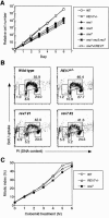
Growth and cell cycle properties of wild-type and rev mutant DT40 cells. (A) Growth curves of WT and mutant DT40 cells. Each value represents the mean of the results from two independent clones for each genotype. (B) Representative cell cycle distribution of the indicated cell cultures as measured by bromodeoxyuridine (BrdU) incorporation and DNA content in flow cytometric analysis. Each of the gates at the upper half, lower left, and lower right and the leftmost gate correspond to cells incorporating BrdU (∼S phase), G1 cells, G2/M cells, and sub-G1 cells, respectively. Numbers show the percentage of cells falling in each gate. (C) Normal spindle assembly checkpoint in rev7 cells. Cells in mitosis were identified by costaining with PI and antibody to phosphorylated histone H3, and the percentages of the mitotic cells are shown as the mitotic index. The period of chase indicates time after addition of 0.1 μg/ml of Colcemid.
FIG. 3.
FIG. 3.
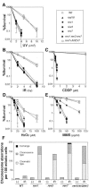
Sensitivities of rev single mutants and rev1 rev3 rev7 cells to killing by various DNA-damaging agents. Results of the colony survival assay after treatment with (A) UV light (254 nm), (B) γ rays (137Cs), (C) cisplatin (CDDP), (D) H2O2, and (E) methyl methanesulfonate (MMS) are indicated. The symbols indicating each genotype, shown at the right top of panels, represent the mean values of at least three independent experiments. Each error bar shows the standard deviation of the mean. The doses of genotoxic agents are displayed on the x axis on a linear scale, and the percentile fractions of surviving colonies are displayed on the y axis on a logarithmic scale. Plating efficiencies of wild-type and mutant cells were ∼80% in the UV, IR, CDDP, and H2O2 assays and were reduced to ∼50% in the MMS assay because of 1 h of incubation in serum-free medium. (F) Level of chromosomal aberrations induced by IR. For each preparation, 100 mitotic cells were analyzed at 3 h after 2-Gy γ-ray (137Cs) irradiation. (−) indicates spontaneous aberrations.
FIG. 4.
FIG. 4.
Level of SCE in asynchronous populations of rev mutant cells. Distribution of spontaneous and 4-NQO-induced SCEs from indicated cells. Solid bars, spontaneous SCEs; shaded bars, SCEs induced by 0.2 ng/ml of 4-NQO. Mean/median numbers of SCEs are shown in parentheses at the top right in each panel. Cells were incubated with bromodeoxyuridine (BrdU) for two cell cycle periods and treated with Colcemid for the last 2 h to enrich mitotic cells. For each preparation, 100 mitotic cells were analyzed. Note that the rev1 mutant was from reference . The Mann-Whitney U test indicated WT < rev1rev3rev7 < rev1 = rev7 = rev3 < rev1 rev3 rev7+cREV1 in spontaneous SCE level and WT = rev1 rev3 rev7 < rev3 = rev1 = rev7 = rev1 rev3 rev7+cREV1 in 4-NQO-induced SCE level (=, not significantly different; <, significantly lower [P < 0.002]).
FIG. 5.
FIG. 5.
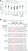
Fluctuation analysis of the generation frequency of sIgM gain revertants. (A) The abundance of sIgM gain variants was determined in 30 parallel cultures derived from single sIgM-negative parental cells after clonal expansion (4 weeks); median percentages are noted below each data set and are indicated by the solid line. The spectra of sIgM gain are indicated on a logarithmic scale. P values calculated by nonparametric statistical analysis (Mann-Whitney U test) between wild-type and mutant DT40 cells are shown. (B) The representative gene conversion tract spectra from sIgM-positive revertants in the wild type and three rev1 mutants are shown on the left as the number of sIgM revertants by gene conversion event/by non-gene conversion event. Each horizontal line represents the rearranged VJλ segment (427 bp) with gene conversion tracts (red lines) and mutations including insertion (arrow), deletion (inverted open triangle), and base substitution (lollipop shape). The parental clone of each genotype has the same frameshift mutation (arrow with G) within this segment. A putative pseudo-V donor is shown on the right of each spectrum with the number of subclones.
FIG. 6.
FIG. 6.
Schematic model of the roles of vertebrate Rev molecules in DNA damage response and HR. Rev1 cooperates with a Rev3-Rev7 complex (i.e., Polζ) as a functional unit in translesion synthesis (TLS) and HR-mediated double-strand break repair (DSBR) for DNA damage response. On the other hand, Rev1 plays a role independent of Polζ in Ig gene conversion, an intragenic HR reaction. In this reaction, Rev1 might cooperate with other Y-family polymerases like Polκ, Polι, or Polη. Rev1 may also act independently of the Rev3-Rev7 complex in the formation of SCE. Thus, Rev1 may regulate two pathways: DNA damage response and intragenic HR.

References

    1. Baynton, K., A. Bresson-Roy, and R. P. Fuchs. 1999. Distinct roles for Rev1p and Rev7p during translesion synthesis in Saccharomyces cerevisiae. Mol. Microbiol. 34:124-133. - PubMed
    1. Bemark, M., A. A. Khamlichi, S. L. Davies, and M. S. Neuberger. 2000. Disruption of mouse polymerase zeta (Rev3) leads to embryonic lethality and impairs blastocyst development in vitro. Curr. Biol. 10:1213-1216. - PubMed
    1. Bezzubova, O., A. Silbergleit, Y. Yamaguchi-Iwai, S. Takeda, and J. M. Buerstedde. 1997. Reduced X-ray resistance and homologous recombination frequencies in a RAD54−/− mutant of the chicken DT40 cell line. Cell 89:185-193. - PubMed
    1. Broomfield, S., T. Hryciw, and W. Xiao. 2001. DNA postreplication repair and mutagenesis in Saccharomyces cerevisiae. Mutat. Res. 486:167-184. - PubMed
    1. Buerstedde, J. M., C. A. Reynaud, E. H. Humphries, W. Olson, D. L. Ewert, and J. C. Weill. 1990. Light chain gene conversion continues at high rate in an ALV-induced cell line. EMBO J. 9:921-927. - PMC - PubMed

Publication types

Substances