. 2005 Jul;25(14):6112-22.
doi: 10.1128/MCB.25.14.6112-6122.2005.

Structural dynamics of alpha-actinin-vinculin interactions

Affiliations

Structural dynamics of alpha-actinin-vinculin interactions

Philippe R J Bois et al. Mol Cell Biol. 2005 Jul.

Erratum in

  • Mol Cell Biol. 2007 Aug;27(15):5606

Abstract

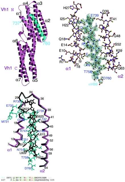
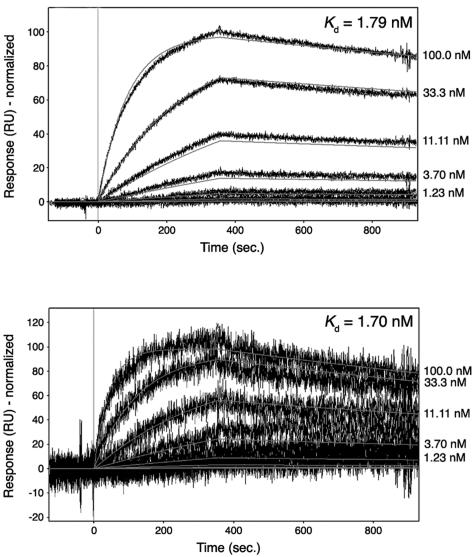
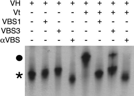
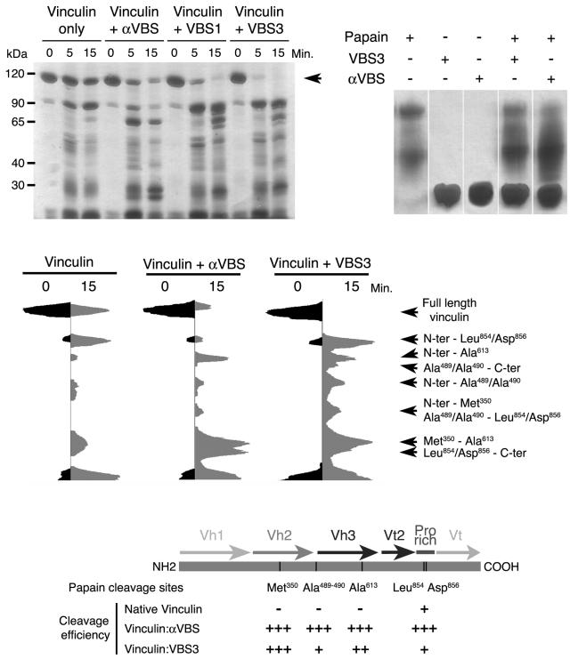
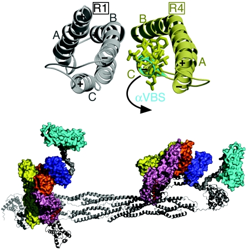
Alpha-actinin and vinculin orchestrate reorganization of the actin cytoskeleton following the formation of adhesion junctions. alpha-Actinin interacts with vinculin through the binding of an alpha-helix (alphaVBS) present within the R4 spectrin repeat of its central rod domain to vinculin's N-terminal seven-helical bundle domain (Vh1). The Vh1:alphaVBS structure suggests that alphaVBS first unravels from its buried location in the triple-helical R4 repeat to allow it to bind to vinculin. alphaVBS binding then induces novel conformational changes in the N-terminal helical bundle of Vh1, which disrupt its intramolecular association with vinculin's tail domain and which differ from the alterations in Vh1 provoked by the binding of talin. Surprisingly, alphaVBS binds to Vh1 in an inverted orientation compared to the binding of talin's VBSs to vinculin. Importantly, the binding of alphaVBS and talin's VBSs to vinculin's Vh1 domain appear to also trigger distinct conformational changes in full-length vinculin, opening up distant regions that are buried in the inactive molecule. The data suggest a model where vinculin's Vh1 domain acts as a molecular switch that undergoes distinct structural changes provoked by talin and alpha-actinin binding in focal adhesions versus adherens junctions, respectively.


Figures

FIG. 1.
FIG. 1.
Helical bundle conversion of vinculin's Vh1 domain by α-actinin. (Top left) Ribbon representation of vinculin's Vh1 domain (pink) in complex with αVBS (α-actinin residues 737 to 760; cyan). The seven α-helices of the Vh1 domain are labeled (α1 to α7). The N termini of α-helices are marked with a plus sign, while their C termini are indicated with a minus sign, in agreement with the helix dipole moment. The N- and C-terminal helical bundles are labeled (Vh1 N and Vh1 C, respectively). (Top right) Final omit electron density map for αVBS (gray bonds, blue labels) bound to helices α1 and α2 of Vh1 (pink bonds, black labels). The contour level of the electron density map is 2σ, and the resolution is 1.8 Å. αVBS refined to a temperature factor of 36.2 Å2. (Bottom left) Top panel, Cα-trace superposition of helices α1 and α2 of the Vh1-talin-VBS1 structure (gray) onto the Vh1:αVBS structure (pink). Talin-VBS1 bonds are shown in black, and αVBS bonds are shown in cyan. Some Cα positions are numbered, and specific peptide residues are labeled (talin-VBS1 in black and αVBS in blue). While the 2.7-Å Vh1-talin-VBS3 (19) structure is very similar to our human 2.4-Å Vh1-talin-VBS1 structure (20) and to the chicken 2-Å Vh1-talin-VBS1 structure (33), for clarity only the higher-resolution Vh1-talin-VBS1 structure is superimposed onto our Vh1-αVBS structure, but similar results are obtained when superimposing all known Vh1-talin-VBS structures. Bottom panel, Structure-based amino acid sequence alignment of αVBS in its reverse (C- to N-terminal) orientation onto talin-VBS1. Identical residues are in red, and similar residues are in green.
FIG. 2.
FIG. 2.
Binding affinities of αVBS for vinculin. Biacore surface plasmon resonance was used to measure the affinity of αVBS. Biotinylated Vh1 or full-length human vinculin was captured on a carboxymethyldextran-coated gold surface (CM-5 chip; Biacore). αVBS peptide was injected over the reference and Vh1- or vinculin-immobilized cells in sequence, and release of bound αVBS peptide was determined. Responses were referenced for reference flow cell and blank injections of buffer. Representative sensorgrams are shown. (Top) Affinity of αVBS for vinculin's Vh1 domain (residues 1 to 258). Binding responses were fit using a mass transport model, and the calculated Kd is shown in the inset. (Bottom) Affinity of αVBS for full-length human vinculin (residues 1 to 1066). Binding responses were fit using a simple model and the calculated Kd is shown in the inset. The signal appears noisier for full-length vinculin compared to Vh1 because of the lower levels of vinculin on the surface, resulting in lower binding capacity.
FIG. 3.
FIG. 3.
Binding of talin-VBS1, talin-VBS3, or αVBS peptides to vinculin's head domain (VH, human residues 1 to 780) is sufficient to displace Vt from preexisting VH-Vt complexes. Native polyacrylamide gel electrophoresis analysis of free VH (lane 1) and of VH bound to talin-VBS1 (lane 2), talin-VBS3 (lane3), αVBS (lane 4), or Vt (human vinculin residues 879 to 1066; lane 5) is shown. Free VH is indicated with an asterisk. Talin-VBS1, talin-VBS3, and αVBS were then added to preexisting VH-Vt complexes (circle) at a fourfold molar excess and displaced Vt to form the respective VH-talin-VBS1 (lane 6), VH-talin-VBS3 (lane 7), or VH-αVBS (lane 8). Free Vt is not visible in native gels because of its basic pI.
FIG. 4.
FIG. 4.
αVBS and talin's VBSs provoke unique conformational alterations in full-length vinculin. (Top left) αVBS and talin's VBS1 and VBS3 alter the sensitivity of human full-length vinculin to papain cleavage. Full-length human vinculin protein was left untreated or was bound with αVBS, talin-VBS1, or talin-VBS3 peptides for 15 min. The proteins were then treated with papain, and at the indicated intervals digestion products of vinculin were analyzed by polyacrylamide gel electrophoresis and silver staining of the gels. αVBS-, talin-VBS1-, and talin-VBS3-bound vinculin were much more susceptible to papain cleavage at Met-350 and Ala-613, indicating that binding of all of these VBSs exposes regions buried in the inactive vinculin structure (5). The arrow indicates full-length (uncleaved) vinculin. (Top right) αVBS and talin's VBS1 and VBS3 are not substrates of papain. αVBS, talin-VBS1, or talin-VBS3 peptides were treated with papain and after 15 min were analyzed by polyacrylamide gel electrophoresis and silver staining of the gels. αVBS, talin-VBS1, and talin-VBS3 were not cleaved by papain. Further, there were no obvious effects of these VBSs on papain activity, as levels of self-digestion products of papain (which were very minor) were essentially unaffected. (Middle) Densitometric scans of papain cleavage products of unbound vinculin or of αVBS- or talin-VBS3-bound vinculin. The cleavage sites were identified by mass spectrometry, N-terminal sequencing, and immunoblotting with anti-His tag antibody (data not shown). The scans of talin-VBS1- andtalin-VBS3-bound vinculin were essentially equivalent (data not shown). By contrast, note the very distinct patterns of papain digestion products of αVBS- versus talin-VBS3-bound vinculin, in particular at Ala-490, which resides in the interface of the Vh2-Vt interaction in inactive vinculin (5) and which is especially prominent in talin-VBS3-bound vinculin. (Bottom) Schematic representation of the locations of the papain cleavage sites and their cleavage efficiency in inactive and αVBS- or talin-VBS3-bound vinculin. Plus signs indicate the relative efficiency of papain cleavage at the indicated sites (e.g., at Met-350 and Ala-613, papain cleavage was 16-fold more efficient in αVBS-bound vinculin than in inactive vinculin). The results shown are representative of those from eight independent experiments.
FIG. 5.
FIG. 5.
Proposed movements of α-actinin's VBS that allow binding to vinculin. (Top) Superposition of α-actinin's vinculin binding site as seen in the native α-actinin rod crystal structure (40) (PDBID 1HCI; gray and yellow) onto αVBS as when bound to the N-terminal helical bundle of Vh1 (cyan). In the dimer of the α-actinin rod, the C-terminal spectrin repeat that contains αVBS (R4, yellow) of one protomer interacts with the N-terminal spectrin repeat (R1, gray) of the other protomer. Residues involved in binding of αVBS to vinculin are shown. In α-actinin's native state, these residues are oriented to the inside of the hydrophobic R4 repeat. The proposed movement that occurs to allow the binding of αVBS to Vh1 is indicated. (Bottom) Model for the binding of the full-length α-actinin dimer (ribbon drawing) to two full-length vinculin molecules (surface representation). The αVBS helix (red) within the R4 spectrin repeat of α-actinin's rod (gray) swings out from its packed position in the α-actinin dimer to expose its hydrophobic face and interact with the N-terminal helical bundle of the Vh1 domain of vinculin (pink). Based on the full-length α-actinin cryo-electron microscopy structure (29) (PDBID 1SJJ), the proposed movements of αVBS also move the C-terminal EF hand domain (black) without steric hindrance. αVBS binding displaces the five-helical bundle of Vt (cyan), which is connected to a four-helical bundle, Vt2 (dark blue), by a proline-rich linker (gray). The two other seven-helical bundles of vinculin, Vh2 (yellow) and Vh3 (orange), are also shown.

Comment in

References

    1. Altmann, S. M., R. G. Grunberg, P. F. Lenne, J. Ylanne, A. Raae, K. Herbert, M. Saraste, M. Nilges, and J. K. Horber. 2002. Pathways and intermediates in forced unfolding of spectrin repeats. Structure 10:1085-1096. - PubMed
    1. Bakolitsa, C., D. M. Cohen, L. A. Bankston, A. A. Bobkov, G. W. Cadwell, L. Jennings, D. R. Critchley, S. W. Craig, and R. C. Liddington. 2004. Structural basis for vinculin activation at sites of cell adhesion. Nature 430:583-586. - PubMed
    1. Bakolitsa, C., J. M. de Pereda, C. R. Bagshaw, D. R. Critchley, and R. C. Liddington. 1999. Crystal structure of the vinculin tail suggests a pathway for activation. Cell 99:603-613. - PubMed
    1. Bass, M. D., B. J. Smith, S. A. Prigent, and D. R. Critchley. 1999. Talin contains three similar vinculin-binding sites predicted to form an amphipathic helix. Biochem. J. 341:257-263. - PMC - PubMed
    1. Borgon, R. A., C. Vonrhein, G. Bricogne, P. R. J. Bois, and T. Izard. 2004. Crystal structure of human vinculin. Structure 12:1189-1197. - PubMed

Publication types