. 2005 Jul;25(14):6199-210.
doi: 10.1128/MCB.25.14.6199-6210.2005.

Loss of histochemical identity in mast cells lacking carboxypeptidase A

Affiliations

Loss of histochemical identity in mast cells lacking carboxypeptidase A

Thorsten B Feyerabend et al. Mol Cell Biol. 2005 Jul.

Abstract

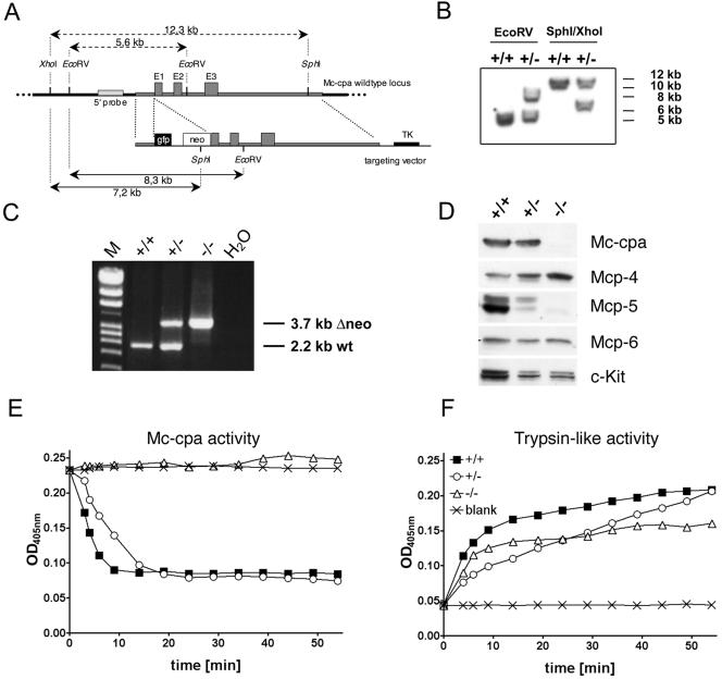
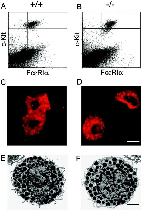
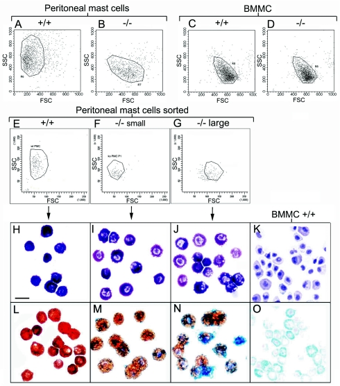
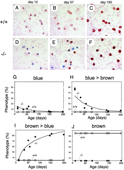
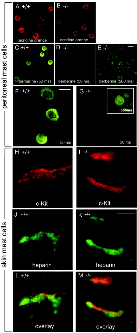
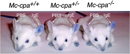
Mast cell carboxypeptidase A (Mc-cpa) is a highly conserved secretory granule protease. The onset of expression in mast cell progenitors and lineage specificity suggest an important role for Mc-cpa in mast cells. To address the function of Mc-cpa, we generated Mc-cpa-null mice. Mc-cpa-/- mast cells lacked carboxypeptidase activity, revealing that Mc-cpa is a nonredundant enzyme. While Mc-cpa-/- peritoneal mast cells were ultrastructurally normal and synthesized normal amounts of heparin, they displayed striking histochemical and biochemical hallmarks of immature mast cells. Wild-type peritoneal mast cells had a mature phenotype characterized by differential histochemical staining with proteoglycan-reactive dyes (cells do not stain with alcian blue but stain with safranin and with berberine) and a high side scatter to forward scatter ratio by flow cytometry and were detergent resistant. In contrast, Mc-cpa-/- peritoneal mast cells, like immature bone marrow-derived cultured mast cells, stained with alcian blue normally or weakly and either did not stain with safranin and berberine or stained weakly, had a low side scatter to forward scatter ratio, and were detergent sensitive. This phenotype was partially ameliorated with age. Thus, histochemistry and flow cytometry, commonly used to measure mast cell maturation, deviated from morphology in Mc-cpa-/- mice. The Mc-cpa-/- mast cell phenotype was not associated with defects in degranulation in vitro or passive cutaneous anaphylaxis in vivo. Collectively, Mc-cpa plays a crucial role for the generation of phenotypically mature mast cells.


Figures

FIG. 1.
FIG. 1.
Generation of Mc-cpa/ mice and protein and enzyme analyses of Mc-cpa/ mast cells. (A) The targeting construct included a short 5′ arm, the enhanced green fluorescent protein gene (gfp), the neomycin resistance gene (neo), a long 3′ arm including exons 1 (from nucleotide +18 on), 2 and 3, and the thymidine kinase (TK) gene. (B) Southern blot of Mc-cpa+/+ (+/+) and Mc-cpa+/ (+/−) ES cell DNA digested with EcoRV or SphI/XhoI. The 5′ probe and expected fragment lengths are shown in panel A. (C) Mc-cpa+/+, Mc-cpa+/, and Mc-cpa/ mice were identified by single 2.2-kb PCR bands, 2.2-kb and 3.7-kb PCR bands, or single 3.7-kb PCR bands. Lane M contains molecular size markers. wt, wild type. (D) Western blot analysis of Mc-cpa+/+, Mc-cpa+/, and Mc-cpa/ mast cells shows the absence of Mc-cpa and Mcp-5 proteins in Mc-cpa/ mast cells. Mcp-4 is upregulated, and Mcp-6 is unaltered. (E and F) Lysates of Mc-cpa+/+ (▪), Mc-cpa+/ (○), and Mc-cpa/ (▵) peritoneal mast cells or the blank control (×) were analyzed for carboxypeptidase activity (E) and trypsin-like activity (F) by test substrates (15). Mc-cpa/ mast cells lacked Mc-cpa activity (E). Cells from all genotypes had trypsin-like activity (F). OD405nm, optical density at 405 nm.
FIG. 2.
FIG. 2.
Mc-cpa/ mast cells are normal with regard to staining for c-Kit and FcɛRI expression, acridine orange staining, and transmission electron microscopy. Peritoneal mast cells from Mc-cpa+/+ (+/+) (A, C, and E) and Mc-cpa/ (−/−) (B, D, and F) mice were analyzed by flow cytometry for c-Kit and FcɛRI expression (A and B) (for relative and absolute mast cell frequencies, see Table 1), by acridine orange staining showing acidic granules (C and D), and by electron microscopy to reveal their ultrastructure (E and F). The scale bars in panel D (12 μm) and panel F (8.5 μm) apply to panels C and D and panels E and F, respectively.
FIG. 3.
FIG. 3.
Mc-cpa/ peritoneal mast cells resemble immature BMMC and possess a mast cell progenitor phenotype. Forward (FSC) and side scatter (SSC) were determined by flow cytometry for Mc-cpa+/+ (+/+) (A, C, and E) and Mc-cpa/ (−/−) (B, D, F, and G) peritoneal mast cells (A, B, E, F, and G) and immature BMMC (C and D). Peritoneal mast cells from Mc-cpa/ mice were separated by cell sorting into small (F) and large (G) subpopulations. Cytospins from the indicated peritoneal mast cell populations were stained with toluidine blue (H to J) and with alcian blue and safranin (L to N) and compared to BMMC (K and O). The scale bar in panel H (20 μm) applies to panels H to O. wt, +/+; ko, −/−.
FIG. 4.
FIG. 4.
Age-dependent block in mast cell development in Mc-cpa/ mice. Peritoneal cells were isolated from wild-type (Mc-cpa+/+ [+/+]) (A to C) and Mc-cpa/ (−/−) (D to F) mice at the indicated age. Cells were cytospun and stained by alcian blue and safranin. Relative proportions of AB+++ S (blue) (G), AB+++ S+ (blue > brown) (H), AB+ S+++ (brown > blue) (I), and AB S+++ (brown) (J) staining (+++, strong staining; +, staining; −, no staining) are shown as a function of time.
FIG. 5.
FIG. 5.
Berberine fluorescence and immunofluorescence analysis for heparin expression in Mc-cpa/ mast cells. Peritoneal cells from wild-type (Mc-cpa+/+ [+/+]) (A, C, and F), and Mc-cpa/ (−/−) (B, D, E, and G) mice were stained with acridine orange to visualize mast cells (A and B) and with berberine sulfate to visualize heparin-containing mast cell granules (C to G). Mc-cpa/ peritoneal mast cells lacked berberine reactivity. Faint nuclear/perinuclear berberine fluorescence was detectable after 10-fold-longer exposure of the film to the fluorescence in Mc-cpa/ mast cells (G). Skin sections from wild-type (H, J, and L) and Mc-cpa/ (I, K, and M) mice were analyzed by immunofluorescence analysis using antiheparin and anti-c-Kit antibodies for the presence of cells expressing c-Kit (H and I), and heparin (J and K). Overlays are shown in panels L and M. All mast cells coexpressed c-Kit and heparin. The scale bars in panels E (20 μm), F (15 μm), and K (10 μm) apply to panels A to E, F and G, and H to M, respectively.
FIG. 6.
FIG. 6.
(A) Heparin biosynthesis and secretion in Mc-cpa+/+ and Mc-cpa/ mast cells. Sorted peritoneal (a) or cultured BM-derived (b) mast cells were metabolically labeled by overnight culture in medium containing [35S]sulfate. Percent radioactivity in counts per minute (cpm) recovered in either the lysis fraction (L1) or salt extraction fraction (L2) is shown for Mc-cpa+/+ (+/+) (solid bars) and Mc-cpa/ (−/−) (open bars) mast cells. Proteoglycans were further fractionated by ion-exchange chromatography and LiCl gradient elution (c). The left and right peaks correspond to chondroitin sulfate and heparin, respectively (10). The heparin peaks disappeared after treatment of the material with HNO2 prior to ion-exchange chromatography (d). The left y axes in panels c and d show percent counts per minute, and the right y axes show conductivity of the LiCl gradient in milli-Siemens per centimeter. Mast cells were pulse-chase-labeled with 35S. There was no spontaneous release into the supernatant (sup), but the material was retained in fractions L1 and L2 (e). w/o, without ionomycin. Following ionomycin (iono) stimulation, mast cells released labeled proteoglycans into the supernatant (sup) (f). (B) Mc-cpa/ mast cells are highly detergent sensitive. Peritoneal mast cells from Mc-cpa+/+ (a to d) and Mc-cpa/ (e to h) mice were cytocentrifuged in the absence (without [w/o]) (a and e) or presence of increasing amounts of Triton X-100(0.016% [b and f], 0.02% [c and g], and 0.5% [d and h]) and stained with toluidine blue. Wild-type mast cells withstood this treatment up to 0.5% Triton X-100 (d) but Mc-cpa/ mast cells showed “holes” at low detergent concentrations (f) and disintegrated entirely at higher concentrations (g and h). The arrowheads in panel h point to the remaining nuclei. The scale bar in panel E (20 μm) applies to panels a to h.
FIG. 7.
FIG. 7.
Mc-cpa/ mast cells mount a normal passive cutaneous anaphylaxis reaction. Mc-cpa+/+, Mc-cpa+/, and Mc-cpa/ mice were injected intradermally into the left and right ears with hapten (DNP)-specific IgE or PBS, respectively. On the next day, mice were challenged by intravenous injection of hapten-carrier (DNP-HSA) together with Evan's blue. The PCA reaction is evident by extravasation of the dye in IgE-injected but not in control PBS-injected ears. Mice of all genotypes mounted indistinguishable PCA reactions. One representative mouse of five mice for each genotype are shown.

References

    1. Chen, X. J., and L. Enerback. 1999. Immature peritoneal mast cells in neonatal rats express the CTMC phenotype, as well as functional IgE receptors. APMIS 107:957-965. - PubMed
    1. Cool, D. R., E. Normant, F. Shen, H. C. Chen, L. Pannell, Y. Zhang, and Y. P. Loh. 1997. Carboxypeptidase E is a regulated secretory pathway sorting receptor: genetic obliteration leads to endocrine disorders in Cpe(fat) mice. Cell 88:73-83. - PubMed
    1. Dvorak, A. M. 1987. Procedural guide to specimen handling for the ultrastructural pathology service laboratory. J. Electron Microsc. Tech. 6:255-301.
    1. Dvorak, A. M., R. A. Seder, W. E. Paul, E. S. Morgan, and S. J. Galli. 1994. Effects of interleukin-3 with or without the c-kit ligand, stem cell factor, on the survival and cytoplasmic granule formation of mouse basophils and mast cells in vitro. Am. J. Pathol. 144:160-170. - PMC - PubMed
    1. Eklund, K. K., N. Ghildyal, K. F. Austen, D. S. Friend, V. Schiller, and R. L. Stevens. 1994. Mouse bone marrow-derived mast cells (mBMMC) obtained in vitro from mice that are mast cell-deficient in vivo express the same panel of granule proteases as mBMMC and serosal mast cells from their normal littermates. J. Exp. Med. 180:67-73. - PMC - PubMed

Publication types

LinkOut - more resources