. 2005 Jul;25(14):6235-46.
doi: 10.1128/MCB.25.14.6235-6246.2005.

The oncogenic TLS-ERG fusion protein exerts different effects in hematopoietic cells and fibroblasts

Affiliations

The oncogenic TLS-ERG fusion protein exerts different effects in hematopoietic cells and fibroblasts

Junhui Zou et al. Mol Cell Biol. 2005 Jul.

Abstract

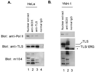
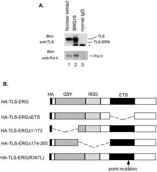
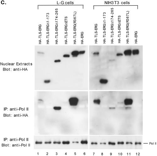
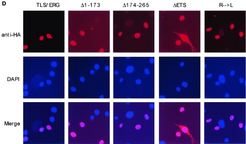
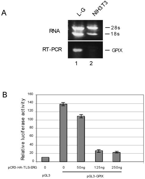
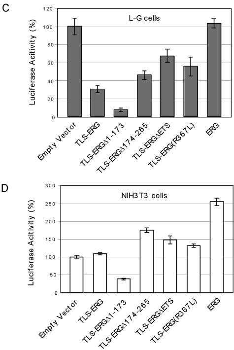
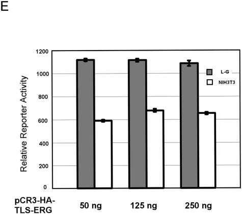
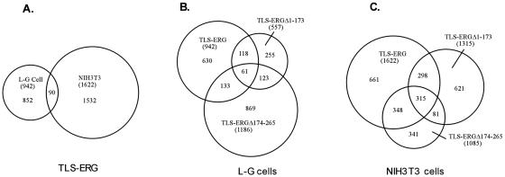
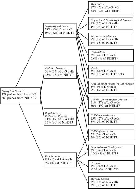
The oncogenic TLS-ERG fusion protein is found in human myeloid leukemia and Ewing's sarcoma as a result of specific chromosomal translocation. To unveil the potential mechanism(s) underlying cellular transformation, we have investigated the effects of TLS-ERG on both gene transcription and RNA splicing. Here we show that the TLS protein forms complexes with RNA polymerase II (Pol II) and the serine-arginine family of splicing factors in vivo. Deletion analysis of TLS-ERG in both mouse L-G myeloid progenitor cells and NIH 3T3 fibroblasts revealed that the RNA Pol II-interacting domain of TLS-ERG resides within the first 173 amino acids. While TLS-ERG repressed expression of the luciferase reporter gene driven by glycoprotein IX promoter in L-G cells but not in NIH 3T3 cells, the fusion protein was able to affect splicing of the E1A reporter in NIH 3T3 cells but not in L-G cells. To identify potential target genes of TLS-ERG, the fusion protein and its mutants were stably expressed in both L-G and NIH 3T3 cells through retroviral transduction. Microarray analysis of RNA samples from these cells showed that TLS-ERG activates two different sets of genes sharing little similarity in the two cell lines. Taken together, these results suggest that the oncogenic TLS-ERG fusion protein transforms hematopoietic cells and fibroblasts via different pathways.


Figures

FIG. 1.
FIG. 1.
Detection of RNA Pol II-TLS-SR protein complexes. (A) HeLa nuclear extract (lane 1) was immunoprecipitated with the mouse monoclonal anti-SC35 (lane 2), a mouse monoclonal antibody against the N-terminal region of TLS (lane 3), or a nonspecific mouse IgG as a negative control (lane 4). The nuclear extract and the immunoprecipitates were blotted with the H5 anti-RNA Pol IIo (top panel), a rabbit polyclonal anti-TLS (middle panel), and the m104 antibody recognizing a family of SR proteins (bottom panel). (B) A similar immunoprecipitation experiment was carried out with the t(16;21) acute leukemia cell line YNH-1 expressing endogenous TLS-ERG. Note the absence of TLS-ERG from the anti-SC35 immunoprecipitate. The position of the IgG band is indicated by an asterisk.
FIG. 2.
FIG. 2.
Interaction of RNA Pol II with the first 173 amino acids of TLS-ERG. (A) YNH-1 nuclear extract (lane 1) was incubated with the anti-Pol II antibody 8WG16 (lane 2) or normal mouse IgG as a negative control (lane 3). The position of the IgG band is indicated by an asterisk. (B) Schematic of HA-TLS-ERG and its mutants with different domains deleted or mutated. HA, hemagglutinin epitope; QSY, glutamine-, serine-, and tyrosine-rich domain; RGG, region with multiple Arg-Gly-Gly repeats; ETS, ets DNA-binding domain. (C) Nuclear extracts from L-G cells (lanes 1 to 6) and NIH 3T3 cells (lanes 7 to 12) expressing HA-TLS-ERG or its mutants were blotted with the 3F10 anti-HA antibody (top panels). The nuclear extracts were incubated with 8WG16, and the immunoprecipitates (IP) were blotted with the anti-HA antibody (middle panels) or the C21 anti-Pol II antibody (bottom panels). (D) NIH 3T3 cells expressing HA-TLS-ERG or its mutants were stained with a Cy3-conjugated anti-HA antibody (top panel), the nuclei were indicated by DAPI staining (middle panel), and the two images were merged to show subcellular localization of the HA-tagged protein.
FIG. 2.
FIG. 2.
Interaction of RNA Pol II with the first 173 amino acids of TLS-ERG. (A) YNH-1 nuclear extract (lane 1) was incubated with the anti-Pol II antibody 8WG16 (lane 2) or normal mouse IgG as a negative control (lane 3). The position of the IgG band is indicated by an asterisk. (B) Schematic of HA-TLS-ERG and its mutants with different domains deleted or mutated. HA, hemagglutinin epitope; QSY, glutamine-, serine-, and tyrosine-rich domain; RGG, region with multiple Arg-Gly-Gly repeats; ETS, ets DNA-binding domain. (C) Nuclear extracts from L-G cells (lanes 1 to 6) and NIH 3T3 cells (lanes 7 to 12) expressing HA-TLS-ERG or its mutants were blotted with the 3F10 anti-HA antibody (top panels). The nuclear extracts were incubated with 8WG16, and the immunoprecipitates (IP) were blotted with the anti-HA antibody (middle panels) or the C21 anti-Pol II antibody (bottom panels). (D) NIH 3T3 cells expressing HA-TLS-ERG or its mutants were stained with a Cy3-conjugated anti-HA antibody (top panel), the nuclei were indicated by DAPI staining (middle panel), and the two images were merged to show subcellular localization of the HA-tagged protein.
FIG. 3.
FIG. 3.
Effects of TLS-ERG and its mutants on GPIX promoter in L-G and NIH 3T3 cells. (A) Expression of the mouse GPIX gene was examined by RT-PCR in L-G (lane 1) and NIH 3T3 (lane 2) cells. (B) pGL3 or pGL3-GPIX firefly luciferase reporter construct (1.5 μg) was introduced into L-G cells with increasing amounts of pCR3-HA-TLS-ERG as indicated. The total amount of the DNA was kept constant by the addition of pCR3 empty vector. (C) pGL3-GPIX (1.5 μg) was cotransfected into L-G cells with 250 ng of pCR3-HA-TLS-ERG construct expressing TLS-ERG or its mutants. (D) pGL3-GPIX (1.5 μg) was cotransfected into NIH 3T3 cells with 250 ng of pCR3-HA-TLS-ERG construct expressing TLS-ERG or its mutants. (E) pGL3-ESET firefly luciferase reporter construct (1.5 μg) was introduced into L-G and NIH 3T3 cells with increasing amounts of pCR3-HA-TLS-ERG as indicated. All transfections were carried out independently at least three times, and the promoter activity was normalized to that of the Renilla luciferase control.
FIG. 3.
FIG. 3.
Effects of TLS-ERG and its mutants on GPIX promoter in L-G and NIH 3T3 cells. (A) Expression of the mouse GPIX gene was examined by RT-PCR in L-G (lane 1) and NIH 3T3 (lane 2) cells. (B) pGL3 or pGL3-GPIX firefly luciferase reporter construct (1.5 μg) was introduced into L-G cells with increasing amounts of pCR3-HA-TLS-ERG as indicated. The total amount of the DNA was kept constant by the addition of pCR3 empty vector. (C) pGL3-GPIX (1.5 μg) was cotransfected into L-G cells with 250 ng of pCR3-HA-TLS-ERG construct expressing TLS-ERG or its mutants. (D) pGL3-GPIX (1.5 μg) was cotransfected into NIH 3T3 cells with 250 ng of pCR3-HA-TLS-ERG construct expressing TLS-ERG or its mutants. (E) pGL3-ESET firefly luciferase reporter construct (1.5 μg) was introduced into L-G and NIH 3T3 cells with increasing amounts of pCR3-HA-TLS-ERG as indicated. All transfections were carried out independently at least three times, and the promoter activity was normalized to that of the Renilla luciferase control.
FIG. 4.
FIG. 4.
Effects of TLS-ERG and its mutants on TASR-mediated E1A pre-mRNA splicing in L-G and NIH 3T3 cells. (A) Diagram of E1A genomic DNA and different splicing isoforms. Numbers indicate individual exons, and dashed lines represent spliced sequences. (B) pCS3-MT-E1A splicing reporter was cotransfected into L-G cells with an empty vector (lane 1) or plasmid expressing Flag-TASR-1 (lanes 2 and 3) or Flag-TASR-2 (lanes 4 and 5) in the presence or absence of TLS-ERG. In NIH 3T3 cells, the pCS3-MT-E1A splicing reporter was cotransfected with an empty vector (lane 6) or plasmids expressing Flag-TASR-1 (lane 7) plus TLS-ERG or its mutants (lanes 8 to 12). In vivo alternative splicing of E1A pre-mRNA was analyzed by an RNase protection assay. Protected E1A RNA fragments are shown on the right, with exons designated by numerals in boxes. Nuclear extracts from the transfected cells were blotted with an anti-Flag antibody to show comparable expression of Flag-tagged TASR proteins (bottom panels).
FIG. 5.
FIG. 5.
Venn diagrams of genes affected by TLS-ERG and its mutants in L-G and NIH 3T3 cells. (A) RNAs from L-G and NIH 3T3 cells stably expressing HA-TLS-ERG were used to analyze gene expression patterns on the Affymetrix GeneChips (mouse genome 430, version 2.0, array). Probe sets that differed from the HA-TLS-ERGΔETS baseline control by at least twofold were selected. The sizes of the Venn circles are proportional to the total number of modulated probe sets. Venn numbers correspond to overlapped probe sets between L-G and NIH 3T3 cells. RNAs from L-G (B) and NIH 3T3 (C) cells stably expressing HA-TLS-ERG, HA-TLS-ERGΔ1-173, or HA-TLS-ERGΔ174-265 were analyzed by the mouse genome 430, version 2.0, array using HA-TLS-ERGΔETS as the baseline control. The total number of probe sets that were affected twofold or higher by each construct is included in parentheses. The number of probe sets shared by the mutants is also indicated.
FIG. 6.
FIG. 6.
Biological processes affected by the transforming HA-TLS-ERG and its mutants in L-G and NIH 3T3 cells. One hundred seventy-nine probe sets shared by the transforming TLS-ERG and TLS-ERGΔ1-173 in L-G cells and 663 probe sets shared by the transforming TLS-ERG and TLS-ERGΔ174-265 in NIH 3T3 cells were analyzed using the Affymetrix GO browser. After the analysis, subgroups of the probe sets (with the number in parentheses) were assigned to the specific biological processes indicated in italics. As a gene product can potentially be involved in more than one biological process, a probe set may thus be assigned to more than one subgroup. Some of the probe sets were not assigned due to lack of information regarding their gene products.

Similar articles

Cited by

  • TASR-1 regulates alternative splicing of collagen genes in chondrogenic cells.
    Matsushita H, Blackburn ML, Klineberg E, Zielinska-Kwiatkowska A, Bolander ME, Sarkar G, Suva LJ, Chansky HA, Yang L. Matsushita H, et al. Biochem Biophys Res Commun. 2007 May 4;356(2):411-7. doi: 10.1016/j.bbrc.2007.02.159. Epub 2007 Mar 9. Biochem Biophys Res Commun. 2007. PMID: 17367759 Free PMC article.
  • Modeling initiation of Ewing sarcoma in human neural crest cells.
    von Levetzow C, Jiang X, Gwye Y, von Levetzow G, Hung L, Cooper A, Hsu JH, Lawlor ER. von Levetzow C, et al. PLoS One. 2011 Apr 29;6(4):e19305. doi: 10.1371/journal.pone.0019305. PLoS One. 2011. PMID: 21559395 Free PMC article.
  • ERG deregulation induces PIM1 over-expression and aneuploidy in prostate epithelial cells.
    Magistroni V, Mologni L, Sanselicio S, Reid JF, Redaelli S, Piazza R, Viltadi M, Bovo G, Strada G, Grasso M, Gariboldi M, Gambacorti-Passerini C. Magistroni V, et al. PLoS One. 2011;6(11):e28162. doi: 10.1371/journal.pone.0028162. Epub 2011 Nov 30. PLoS One. 2011. PMID: 22140532 Free PMC article.
  • A causal role for ERG in neoplastic transformation of prostate epithelium.
    Klezovitch O, Risk M, Coleman I, Lucas JM, Null M, True LD, Nelson PS, Vasioukhin V. Klezovitch O, et al. Proc Natl Acad Sci U S A. 2008 Feb 12;105(6):2105-10. doi: 10.1073/pnas.0711711105. Epub 2008 Feb 1. Proc Natl Acad Sci U S A. 2008. PMID: 18245377 Free PMC article.
  • Deregulation of DUX4 and ERG in acute lymphoblastic leukemia.
    Zhang J, McCastlain K, Yoshihara H, Xu B, Chang Y, Churchman ML, Wu G, Li Y, Wei L, Iacobucci I, Liu Y, Qu C, Wen J, Edmonson M, Payne-Turner D, Kaufmann KB, Takayanagi SI, Wienholds E, Waanders E, Ntziachristos P, Bakogianni S, Wang J, Aifantis I, Roberts KG, Ma J, Song G, Easton J, Mulder HL, Chen X, Newman S, Ma X, Rusch M, Gupta P, Boggs K, Vadodaria B, Dalton J, Liu Y, Valentine ML, Ding L, Lu C, Fulton RS, Fulton L, Tabib Y, Ochoa K, Devidas M, Pei D, Cheng C, Yang J, Evans WE, Relling MV, Pui CH, Jeha S, Harvey RC, Chen IL, Willman CL, Marcucci G, Bloomfield CD, Kohlschmidt J, Mrózek K, Paietta E, Tallman MS, Stock W, Foster MC, Racevskis J, Rowe JM, Luger S, Kornblau SM, Shurtleff SA, Raimondi SC, Mardis ER, Wilson RK, Dick JE, Hunger SP, Loh ML, Downing JR, Mullighan CG; St. Jude Children's Research Hospital–Washington University Pediatric Cancer Genome Project. Zhang J, et al. Nat Genet. 2016 Dec;48(12):1481-1489. doi: 10.1038/ng.3691. Epub 2016 Oct 24. Nat Genet. 2016. PMID: 27776115 Free PMC article.

References

    1. Arvand, A., H. Bastians, S. M. Welford, A. D. Thompson, J. V. Ruderman, and C. T. Denny. 1998. EWS/FLI1 up regulates mE2-C, a cyclin-selective ubiquitin conjugating enzyme involved in cyclin B destruction. Oncogene 17:2039-2045. - PubMed
    1. Bastian, L. S., M. Yagi, C. Chan, and G. J. Roth. 1996. Analysis of the megakaryocyte glycoprotein IX promoter identifies positive and negative regulatory domains and functional GATA and Ets sites. J. Biol. Chem. 271:18554-18560. - PubMed
    1. Bertolotti, A., Y. Lutz, D. J. Heard, P. Chambon, and L. Tora. 1996. hTAF(II)68, a novel RNA/ssDNA-binding protein with homology to the pro- oncoproteins TLS/FUS and EWS is associated with both TFIID and RNA polymerase II. EMBO J. 15:5022-5031. - PMC - PubMed
    1. Blackburn, M. L., H. A. Chansky, A. Zielinska-Kwiatkowska, Y. Matsui, and L. Yang. 2003. Genomic structure and expression of the mouse ESET gene encoding an ERG-associated histone methyltransferase with a SET domain. Biochim. Biophys. Acta 1629:8-14. - PubMed
    1. Caceres, J. F., S. Stamm, D. M. Helfman, and A. R. Krainer. 1994. Regulation of alternative splicing in vivo by overexpression of antagonistic splicing factors. Science 265:1706-1709. - PubMed

Publication types