

Neurologic recovery according to early magnetic resonance imaging findings in traumatic cervical spinal cord injuries
- PMID: 15988810
- PMCID: PMC2815815
- DOI: 10.3349/ymj.2005.46.3.379
Neurologic recovery according to early magnetic resonance imaging findings in traumatic cervical spinal cord injuries
Abstract
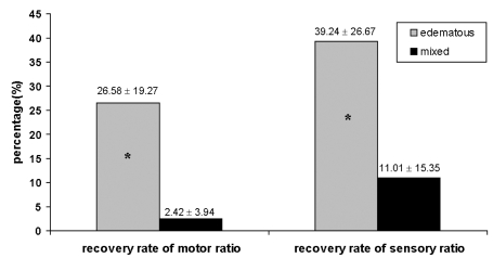
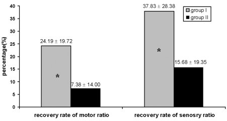
The aim of this study was to determine the usefulness of early magnetic resonance imaging findings in predicting neurologic recovery at or below the injured level in traumatic cervical spinal cord injuries. Thirty patients with traumatic cervical spinal cord injuries were included. All of the patients received a magnetic resonance imaging and a neurologic examination in the emergency room, within 7 days of injury and at 6 months following the injury. To quantify neurologic recovery below the injured level, we modified clinical scales, particularly the motor ratio and the sensory ratio. We used the neurologic level to quantify recovery around the injured level. We assessed neurologic recovery according to MRI patterns and lesion extents. The pure hemorrhagic MRI pattern was not observed. In edematous and mixed types, the improvement of neurologic levels was not significantly different. The motor ratio and sensory ratio improved significantly more in edematous type patients than in mixed type patients. Based on MRI lesion extent, the improvement of neurologic levels was not significantly different, and motor ratio and sensory ratio improved significantly more in those with one or two segments involved than in those with more than two segments involved. In conclusion, early MRI pattern and lesion extent after traumatic cervical spinal cord injury may provide important information to help predict neurologic recovery, especially below the injured level.
Figures
Similar articles
-
Predictors of neurologic recovery in acute central cervical cord injury with only upper extremity impairment.Spine (Phila Pa 1976). 2002 Aug 1;27(15):1652-8; discussion 1658. doi: 10.1097/00007632-200208010-00011. Spine (Phila Pa 1976). 2002. PMID: 12163727 Clinical Trial.
-
Acute cervical traumatic spinal cord injury: MR imaging findings correlated with neurologic outcome--prospective study with 100 consecutive patients.Radiology. 2007 Jun;243(3):820-7. doi: 10.1148/radiol.2433060583. Epub 2007 Apr 12. Radiology. 2007. PMID: 17431129
-
Predicting neurologic recovery in cervical spinal cord injury with postoperative MR imaging.Spine (Phila Pa 1976). 2006 Mar 1;31(5):554-9. doi: 10.1097/01.brs.0000201274.59427.a4. Spine (Phila Pa 1976). 2006. PMID: 16508551
-
Predicting neurologic recovery in traumatic cervical spinal cord injury.Arch Phys Med Rehabil. 1998 Nov;79(11):1456-66. doi: 10.1016/s0003-9993(98)90244-1. Arch Phys Med Rehabil. 1998. PMID: 9821910 Review.
-
Predicting Outcomes After Spinal Cord Injury.Phys Med Rehabil Clin N Am. 2020 Aug;31(3):331-343. doi: 10.1016/j.pmr.2020.03.003. Epub 2020 May 26. Phys Med Rehabil Clin N Am. 2020. PMID: 32624098 Review.
Cited by
-
Neuroimaging in traumatic spinal cord injury: an evidence-based review for clinical practice and research.J Spinal Cord Med. 2007;30(3):205-14. doi: 10.1080/10790268.2007.11753928. J Spinal Cord Med. 2007. PMID: 17684886 Free PMC article. Review.
-
Model-based parcellation of diffusion MRI of injured spinal cord predicts hand use impairment and recovery in squirrel monkeys.Behav Brain Res. 2024 Feb 29;459:114808. doi: 10.1016/j.bbr.2023.114808. Epub 2023 Dec 10. Behav Brain Res. 2024. PMID: 38081518 Free PMC article.
References
-
- Go BK, DeVivo MJ, Richards JS. The epidemiology of spinal cord injury. In: Stover SI, DeLisa JA, Whiteneck GG, editors. Spinal cord Injury: clinical outcomes from the model systems. 1st ed. Gaithersberg: Aspen Publishers; 1995. pp. 21–51.
-
- Kirshblum SC, O'Connor KC. Predicting neurologic recovery in traumatic cervical spinal cord injury. Arch Phys Med Rehabil. 1998;79:1456–1466. - PubMed
-
- Flanders AE, Spettell CM, Tartaglino LM, Friedman DP, Herbison GJ. Forecasting motor recovery after cervical spinal cord injury: value of MR imaging. Radiology. 1996;201:649–655. - PubMed
-
- York DH, Watts C, Raffensberger M, Spagnolia T, Joyce C. Utilization of somatosensory evoked cortical potentials in spinal cord injury. Prognostic limitations. Spine. 1983;8:832–839. - PubMed
-
- Armin C. Significance of electrophysiological recording in predicting functional recovery of patients with spinal cord injury. Neurorehabil. 1998;10:191–203.
Publication types
MeSH terms
LinkOut - more resources
Full Text Sources
Medical
