

The basophil activation test by flow cytometry: recent developments in clinical studies, standardization and emerging perspectives
- PMID: 15989690
- PMCID: PMC1190199
- DOI: 10.1186/1476-7961-3-9
The basophil activation test by flow cytometry: recent developments in clinical studies, standardization and emerging perspectives
Abstract
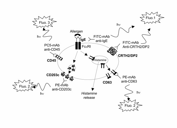
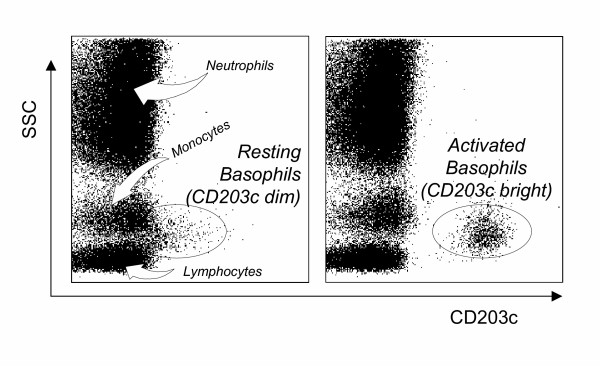
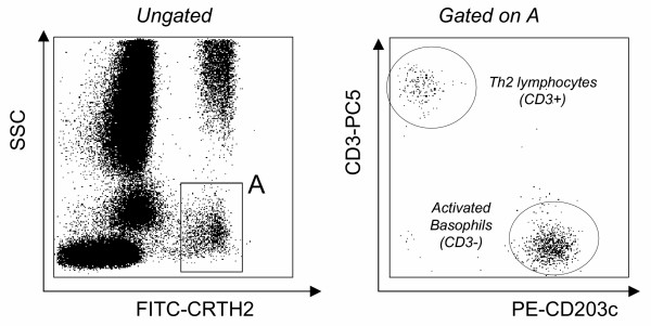
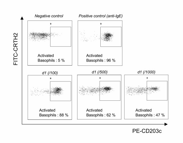
The diagnosis of immediate allergy is mainly based upon an evocative clinical history, positive skin tests (gold standard) and, if available, detection of specific IgE. In some complicated cases, functional in vitro tests are necessary. The general concept of those tests is to mimic in vitro the contact between allergens and circulating basophils. The first approach to basophil functional responses was the histamine release test but this has remained controversial due to insufficient sensitivity and specificity. During recent years an increasing number of studies have demonstrated that flow cytometry is a reliable tool for monitoring basophil activation upon allergen challenge by detecting surface expression of degranulation/activation markers (CD63 or CD203c). This article reviews the recent improvements to the basophil activation test made possible by flow cytometry, focusing on the use of anti-CRTH2/DP2 antibodies for basophil recognition. On the basis of a new triple staining protocol, the basophil activation test has become a standardized tool for in vitro diagnosis of immediate allergy. It is also suitable for pharmacological studies on non-purified human basophils. Multicenter studies are now required for its clinical assessment in large patient populations and to define the cut-off values for clinical decision-making.
Figures
Similar articles
-
Marked improvement of the basophil activation test by detecting CD203c instead of CD63.Clin Exp Allergy. 2003 Feb;33(2):259-65. doi: 10.1046/j.1365-2222.2003.01594.x. Clin Exp Allergy. 2003. PMID: 12580920
-
CD63 expression on basophils as a tool for the diagnosis of pollen-associated food allergy: sensitivity and specificity.Clin Exp Allergy. 2003 May;33(5):607-14. doi: 10.1046/j.1365-2222.2003.01660.x. Clin Exp Allergy. 2003. PMID: 12752589
-
Flow cytometry for basophil activation markers: the measurement of CD203c up-regulation is as reliable as CD63 expression in the diagnosis of cat allergy.J Immunol Methods. 2007 Mar 30;320(1-2):40-8. doi: 10.1016/j.jim.2006.12.002. Epub 2007 Jan 3. J Immunol Methods. 2007. PMID: 17275019
-
The basophil activation test in immediate-type drug allergy.Immunol Allergy Clin North Am. 2009 Aug;29(3):555-66. doi: 10.1016/j.iac.2009.04.011. Immunol Allergy Clin North Am. 2009. PMID: 19563997 Review.
-
Principles, potential, and limitations of ex vivo basophil activation by flow cytometry in allergology: A narrative review.J Allergy Clin Immunol. 2021 Apr;147(4):1143-1153. doi: 10.1016/j.jaci.2020.10.027. Epub 2020 Nov 2. J Allergy Clin Immunol. 2021. PMID: 33152367 Review.
Cited by
-
Behind the scenes with basophils: an emerging therapeutic target.Immunother Adv. 2021 May 19;1(1):ltab008. doi: 10.1093/immadv/ltab008. eCollection 2021 Jan. Immunother Adv. 2021. PMID: 35919744 Free PMC article. Review.
-
Peripheral basophil reactivity, CD203c expression by Cryj1 stimulation, is useful for diagnosing seasonal allergic rhinitis by Japanese cedar pollen.Immun Inflamm Dis. 2015 Sep;3(3):300-8. doi: 10.1002/iid3.69. Epub 2015 Jun 26. Immun Inflamm Dis. 2015. PMID: 26417444 Free PMC article.
-
Current understanding of allergic transfusion reactions: incidence, pathogenesis, laboratory tests, prevention and treatment.Br J Haematol. 2013 Feb;160(4):434-44. doi: 10.1111/bjh.12150. Epub 2012 Dec 6. Br J Haematol. 2013. PMID: 23215650 Free PMC article. Review.
-
Diclofenac induces basophil degranulation without increasing CD63 expression in sensitive patients.Clin Exp Immunol. 2007 Jan;147(1):99-105. doi: 10.1111/j.1365-2249.2006.03265.x. Clin Exp Immunol. 2007. PMID: 17177968 Free PMC article.
-
Carboplatin-induced severe hypersensitivity reaction: role of IgE-dependent basophil activation and FcεRI.Cancer Sci. 2014 Nov;105(11):1472-9. doi: 10.1111/cas.12538. Epub 2014 Nov 5. Cancer Sci. 2014. PMID: 25230301 Free PMC article.
References
-
- Falcone FH, Haas H, Gibbs BF. The human basophil: a new appreciation of its role in immune responses. Blood. 2000;96:4028–38. - PubMed
LinkOut - more resources
Full Text Sources
Other Literature Sources
Miscellaneous
