. 2005 Jul 20;24(14):2579-89.
doi: 10.1038/sj.emboj.7600737. Epub 2005 Jun 30.

The MAP kinase substrate MKS1 is a regulator of plant defense responses

Affiliations

The MAP kinase substrate MKS1 is a regulator of plant defense responses

Erik Andreasson et al. EMBO J. .

Abstract

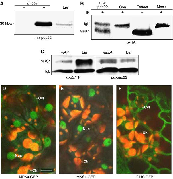
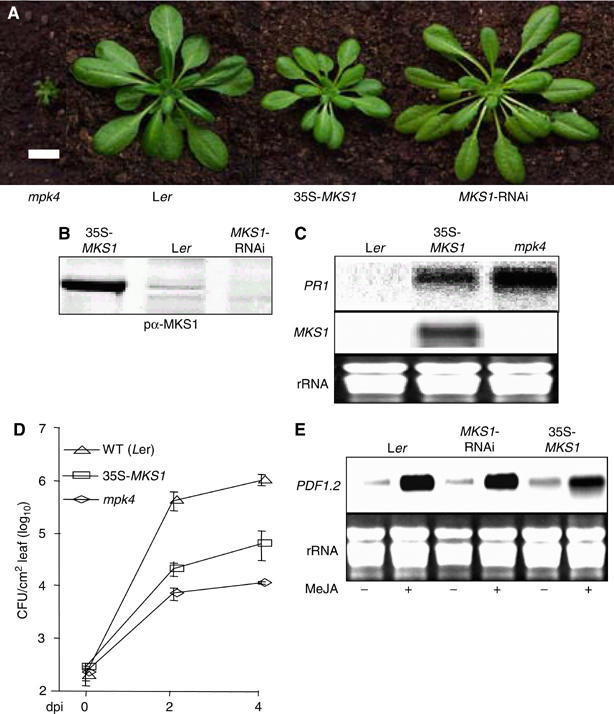
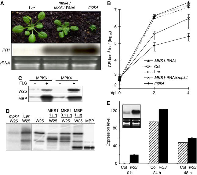
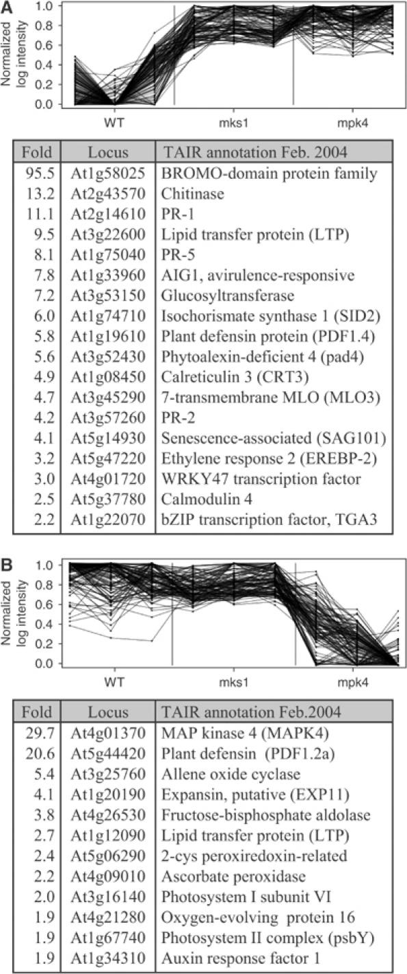
Arabidopsis MAP kinase 4 (MPK4) functions as a regulator of pathogen defense responses, because it is required for both repression of salicylic acid (SA)-dependent resistance and for activation of jasmonate (JA)-dependent defense gene expression. To understand MPK4 signaling mechanisms, we used yeast two-hybrid screening to identify the MPK4 substrate MKS1. Analyses of transgenic plants and genome-wide transcript profiling indicated that MKS1 is required for full SA-dependent resistance in mpk4 mutants, and that overexpression of MKS1 in wild-type plants is sufficient to activate SA-dependent resistance, but does not interfere with induction of a defense gene by JA. Further yeast two-hybrid screening revealed that MKS1 interacts with the WRKY transcription factors WRKY25 and WRKY33. WRKY25 and WRKY33 were shown to be in vitro substrates of MPK4, and a wrky33 knockout mutant was found to exhibit increased expression of the SA-related defense gene PR1. MKS1 may therefore contribute to MPK4-regulated defense activation by coupling the kinase to specific WRKY transcription factors.


Figures

Figure 1
Figure 1
MKS1 interactions and phosphorylation in vitro. (A) MKS1 interactions with MPK4, W25 and W33 used N-terminal 6xHis-tagged MKS1 (His-MKS1) purified from E. coli. 35S-methionine-labeled MPK4, W25 or W33 was incubated without (−) or with (+) nickel-agarose-coupled His-MKS1. Human lamin (Clontech) was a negative control. (B) Left: Phosphorylation of full-length recombinant MKS1, C-terminal MKS1 truncations (C1–C3) and myelin basic protein (MBP) as positive control. HA-tagged MPK4 (HA-MPK4) was immunoprecipitated (IP) from complemented mpk4 transgenic plants. Right: In vitro kinase assays with mutated full-length MKS1 (MKS1 S30A) and mutated C3 (C3 S30A). (C) Phosphorylation of full-length MKS1 and increasing molar ratios of a 22-amino-acid, MKS1-derived peptide, Pep22 (left; Supplementary Figure S1) and the 22-amino-acid flagellin elicitor Flg22 (right).
Figure 2
Figure 2
MKS1 and homologs. DNA and amino-acid sequences of MKS1 (At3g18690) were used to query databases at www.ncbi.nlm.nih.gov/BLAST for proteins similar to MKS1. Protein sequences of selected accessions were aligned at clustalw.genome.jp and identical/similar residues highlighted at www.ch.embnet.org/software/BOX_form.html. Putative MAP kinase phosphorylation sites (S/TP) are underlined. The sequence of the Pep22 peptide is indicated by an overbar. The ends of an N-terminally truncated (N1) and three C-terminally truncated (C1–3) MKS1 versions described in the text are noted above the MKS1 sequence. Putative domains I and II are underlined in the consensus. Species abbreviations are as follows: At: Arabidopsis thaliana; Bo: Brassica oleracea; Gm: Glycine max; St: Solanum tuberosum; Os: Oryza sativa; Nt: Nicotiana tabacum; Zm: Zea mays. Similar plant proteins not aligned here include BM340911, CAD40925, CC613160, CC635639, Al390921, AL138658, T46022, AP004654, AP003260, AC143340 and Arabidopsis At1g21326, At2g41180, At2g44340, At2g42140 and At3g56710 (Sig1; Morikawa et al, 2002).
Figure 3
Figure 3
MKS1 interaction, phosphorylation and localization in vivo. (A) Immunodetection of MKS1 in E. coli extracts before (−) and after (+) IPTG induction, and from extracts of leaves (Ler) by a monoclonal anti-Pep22 antibody (mα-pep22) recognizing a protein of the predicted size of MKS1 (∼28 kDa). (B) Immunodetection of HA-MPK4 by anti-HA antibody (α-HA) in immunoprecipitates from plant extracts with mα-pep22 (lane 1), negative control monoclonal antibody (Con; lane 2) or in mock immunoprecipitation lacking extract (lane 4). MPK4 from plant extracts is shown (raw extract; lane 3). MPK4 and immunoglobin heavy chain (IgH) are indicated. (C) Immunodetection of MKS1 following immunoprecipitation with mα-Pep22 from Ler or mpk4 extracts. MKS1 is detected with an anti-phosphoserine/phosphothreonine antibody (α-pS/TP) and polyclonal anti-Pep22 antibody (pα-pep22). MKS1 and immunoglobin light chain (IgL) are indicated. In planta localization in mesophyll cells of (D) MPK4-GFP, (E) MKS1-GFP and (F) GUS-GFP fusion proteins. Cyt: cytoplasm; Nuc: nuclei; Chl: chloroplast (orange autofluorescent); — 10 μm size bar.
Figure 4
Figure 4
Effects of MKS1 over- and underexpression. (A) Phenotypes of mpk4, wild type (Ler) and transgenic (Col) overexpressing MKS1 (35S-MKS1) or underexpressing MKS1 (MKS1-RNAi). The size bar is 2 cm. (B) Immunodetection with pα-Pep22 antibody of MKS1 in extracts from 35S-MKS1, wild type (Ler) and MKS1-RNAi. (C) RNA blot detection of PR1 mRNA in 35S-MKS1, mpk4 and wild-type Ler. (D) Growth of P. syringae pv. tomato DC3000 in mpk4, 35S-MKS1 and Ler. (E) Detection of PDF1.2 mRNA in wild type (Col), MKS1-RNAi and 35S-MKS1 following MeJA (+) and mock (−) treatments.
Figure 5
Figure 5
Suppression of mpk4 by MKS1-RNAi, WRKY phosphorylation and w33 phenotype. (A) Top: Phenotypes of wild type (Ler), mpk4 carrying MKS1-RNAi (mpk4/MKS1-RNAi) and mpk4. Bottom: Detection of PR1 mRNA in wild type (Ler), mpk4/MKS1-RNAi and mpk4 versus rRNA loading control. (B) Growth of P. syringae pv. tomato DC3000 in wild types Ler and Col, mpk4, MKS1-RNAi and mpk4/MKS1-RNAi. (C) Phosphorylation of full-length W25 by immunoprecipitation of MPK6 or MPK4 from Arabidopsis cells without (−) or with (+) treatment with flagellin. (D) Phosphorylation of N-terminal region of W25 with MPK4 immunoprecipitation from mpk4 or Ler (left) and of W25, MKS1 and MBP with immunoprecipitation of MPK4-HA (right). (E) RNA blot (inset) and real-time PCR detection of PR1 mRNA in Col and w33 before (0 h and inset) or after infiltration with DC3000.
Figure 6
Figure 6
Expression profiles of genes differentially expressed between wild-type, mpk4 and 35S-mks1 plants. Two clusters A and B of significantly differentially expressed genes are shown. (A) Top: Expression profiles of genes in cluster A through triplicates of the three genotypes (x-axis) versus normalized log expression index values. Vertical lines mark the borders between the genotypes. Bottom: List of selected genes from cluster A. (B) as in (A) but for cluster B genes. See Supplementary Figure S1 for a comprehensive list of clustered genes.

References

    1. Asai T, Tena G, Plotnikova J, Willmann MR, Chiu WL, Gomez-Gomez L, Boller T, Ausubel FM, Sheen J (2002) MAP kinase signalling cascade in Arabidopsis innate immunity. Nature 415: 977–983 - PubMed
    1. Baker DA, Mille-Baker B, Wainwright SM, Ish-Horowicz D, Dibb NJ (2001) Mae mediates MAP kinase phosphorylation of Ets transcription factors in Drosophila. Nature 411: 330–334 - PubMed
    1. Bao MZ, Schwartz MA, Cantin GT, Yates JR III, Madhani HD (2004) Pheromone-dependent destruction of the Tec1 transcription factor is required for MAP kinase signaling specificity in yeast. Cell 119: 991–1000 - PubMed
    1. Clarke JD, Liu Y, Klessig DF, Dong X (1998) Uncoupling PR gene expression from NPR1 and bacterial resistance, characterization of the dominant Arabidopsis cpr6-1 mutant. Plant Cell 10: 557–569 - PMC - PubMed
    1. Dangl JL, Jones JD (2001) Plant pathogens and integrated defence responses to infection. Nature 411: 826–833 - PubMed

Publication types