. 2005 Jul;79(14):8812-27.
doi: 10.1128/JVI.79.14.8812-8827.2005.

Evaluating the immunogenicity of a disulfide-stabilized, cleaved, trimeric form of the envelope glycoprotein complex of human immunodeficiency virus type 1

Affiliations

Evaluating the immunogenicity of a disulfide-stabilized, cleaved, trimeric form of the envelope glycoprotein complex of human immunodeficiency virus type 1

Simon Beddows et al. J Virol. 2005 Jul.

Abstract

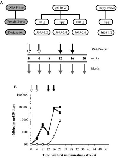
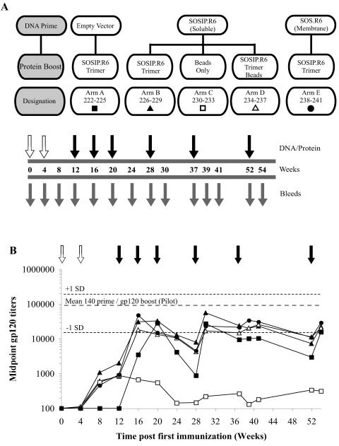
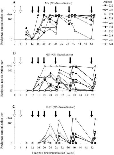
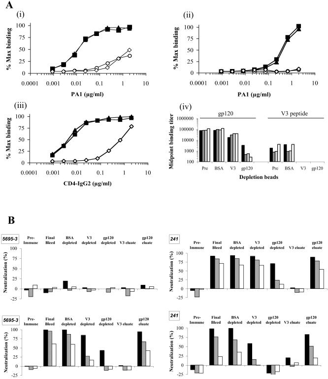
The human immunodeficiency virus type 1 (HIV-1) envelope glycoprotein (Env) complex comprises three gp120 exterior glycoproteins each noncovalently linked to a gp41 transmembrane glycoprotein. Monomeric gp120 proteins can elicit antibodies capable of neutralizing atypically sensitive test viruses in vitro, but these antibodies are ineffective against representative primary isolates and the gp120 vaccines failed to provide protection against HIV-1 transmission in vivo. Alternative approaches to raising neutralizing antibodies are therefore being pursued. Here we report on the antibody responses generated in rabbits against a soluble, cleaved, trimeric form of HIV-1(JR-FL) Env. In this construct, the gp120 and gp41 moieties are covalently linked by an intermolecular disulfide bond (SOS gp140), and an I559P substitution has been added to stabilize gp41-gp41 interactions (SOSIP gp140). We investigated the value of DNA priming and compared the use of membrane-bound and soluble priming antigens and of repeat boosting with soluble and particulate protein antigen. Compared to monomeric gp120, SOSIP gp140 trimers elicited approximately threefold lower titers of anti-gp120 antibodies. Priming with DNA encoding a membrane-bound form of the SOS gp140 protein, followed by several immunizations with soluble SOSIP gp140 trimers, resulted in antibodies capable of neutralizing sensitive strains at high titers. A subset of these sera also neutralized, at lower titers, HIV-1(JR-FL) and some other primary isolates in pseudovirus and/or whole-virus assays. Neutralization of these viruses was immunoglobulin mediated and was predominantly caused by antibodies to gp120 epitopes, but not the V3 region.


Figures

FIG. 1.
FIG. 1.
Env constructs and SOSIP.R6 trimer purification. (A) Schematic of the env DNA constructs used for the priming immunization. Highlighted are the approximate size of the Env protein construct relative to full-length Wt gp160, the inclusion of a Wt (REKR) or enhanced (RRRRRR) cleavage site motif, the approximate position of the SOS cysteine bond (C = C), and the approximate position of the gp41-gp41 trimer-stabilizing mutation (I559P). The C1 to C5 domains of gp120 are also identified. Ecto, TM, and Cyt indicate the positions of the gp41 ectodomain, the membrane-spanning domain, and the cytoplasmic tail, respectively. (B) SDS-PAGE analysis of the purified HIV-1 gp120 and SOSIP.R6 gp140 proteins. The purified proteins, solubilized in Laemmli sample buffer with (reduced) or without (nonreduced) 20 mM dithiothreitol, were resolved on a 4 to 12% Bis-Tris polyacrylamide gradient gel and stained using Coomassie blue. (C) Blue-Native PAGE analysis of the purified HIV-1 gp120 and SOSIP.R6 gp140 proteins. The proteins were resolved on a 4 to 12% Bis-Tris polyacrylamide gradient gel and visualized using Coomassie blue. The values on the left are molecular sizes in kilodaltons.
FIG. 2.
FIG. 2.
Anti-gp120 binding Ab elicited by immunization in the pilot study. (A) The schematic for the pilot study highlights the DNA construct (gp140 Wt, or empty vector) used for priming (open arrows), the dose of gp120 protein (10, 30, or 100 μg) used for each of the protein boosts (filled arrows), and the serum collection times (grey arrows). (B) Generation of anti-gp120 Ab in the pilot study. Rabbits were primed with 1 mg of pPPI4 expressing either codon-optimized soluble gp140 Wt Env (filled symbols) or the empty-vector control (open squares) and then boosted with 10 μg (filled triangles), 30 μg (filled squares), or 100 μg (filled circles) of soluble gp120 at the times indicated. The anti-gp120 Ab responses were measured by ELISA. Each datum point represents the mean (n = 2 animals) midpoint anti-gp120 binding titer for each arm.
FIG. 3.
FIG. 3.
Anti-gp120 binding Ab elicited by SOSIP.R6 immunization. (A) The schematic for the second-stage study highlights the DNA construct [empty vector, SOSIP.R6, or SOS.R6(T)] used for priming (open arrows) the protein construct (SOSIP.R6 trimer, SOSIP.R6 trimer coupled to paramagnetic beads, or empty beads) used for each of the protein boosts (filled arrows), and the serum collection times (grey arrows). The designation numbers for the individual animals in each arm (A to E) are also listed. (B) Rabbits were primed with 1 mg pPPI4 expressing soluble gp140 SOSIP.R6 Env (▴, □, ▵; arms B, C, and D) or membrane-bound SOS.R6 Env (•; arm E) or primed with the empty-vector control (▪; arm A). The animals were then boosted with 30 μg of SOSIP.R6 trimer (▪, ▴, •; arms A, B, and E), 30 μg of SOSIP.R6 trimer coupled to beads (▵; arm D), or empty beads as a control (□; arm C) at the times indicated. The anti-gp120 Ab responses were measured by ELISA. Each datum point represents the mean (n = 4 animals) midpoint anti-gp120 binding titer for each arm. The mean (± standard deviation) anti-gp120 titer in the gp140 Wt DNA-primed and gp120-boosted animals (5695-1 to 5695-6) at week 20 of the pilot study is indicated by the dotted lines for comparison.
FIG. 4.
FIG. 4.
Neutralization of HIV-1JR-FL and HIV-1MN Env-pseudotyped viruses by selected sera. A panel of antisera taken from 10 rabbits immunized with trimeric SOSIP.R6 Env (second-stage study) was assembled to assess the generation of NAb over time. NAb titers against Env-pseudotyped HIV-1MN (A, B) and Env-pseudotyped HIV-1JR-FL (C) were determined at 12 time points over the 54-week time course, using a 50% (A, C) or 90% (B) endpoint. Symbols are used to represent the following groups: empty vector primed and SOSIP.R6 trimer boosted (squares), SOSIP.R6 primed and SOSIP.R6 trimer boosted (triangles), SOSIP.R6 primed and bead-SOSIP.R6 trimer boosted (circles), and membrane-bound SOS.R6(T) primed and SOSIP.R6 trimer boosted (diamonds). The timings of DNA priming (open arrows) and protein boosting (filled arrows) events are indicated. Note that the rabbit sera were not titrated beyond a dilution of 1:160; hence, differences between animals, or the heights of the peaks and depths of the troughs, cannot be determined for values beyond this level.
FIG. 5.
FIG. 5.
Neutralization of HIV-1JR-FL pseudovirus by rabbit antisera is predominantly mediated by non-V3, gp120-directed antibodies. (A) BSA (filled triangles)-, V3 peptide (open circles)-, or gp120 (open diamonds)-coupled cyanogen bromide-Sepharose beads were used to deplete a rabbit preimmune serum pool spiked with V3-specific MAb PA1 (i, ii) or the CD4-IgG2 molecule (iii) before assay by gp120 binding ELISA (i, iii) or V3 peptide ELISA (ii). The nondepleted serum was also assayed (filled squares). (iv) Rabbit final blood drawing sera from the pilot study, 5695-3 (filled bars), and follow-up study, 228 (dark grey bars), 236 (light grey bars), and 241 (open bars), were left untreated (Pre) or depleted using BSA-, V3-, or gp120-coupled beads before determination of midpoint binding titers against gp120 or V3 peptide by ELISA as indicated. (B) Untreated and bead-treated sera 5695-3 and 241 were also used in pseudovirus neutralization assays against HIV-1JR-FL (upper parts) or HIV-1MN (lower parts). Bars represent serum dilutions of 1:10 (filled bars), 1:40 (grey bars), and 1:160 (open bars). Representative data were derived from two or three experiments.

References

    1. Abrahamyan, L. G., R. M. Markosyan, J. P. Moore, F. S. Cohen, and G. B. Melikyan. 2003. Human immunodeficiency virus type 1 Env with an intersubunit disulfide bond engages coreceptors but requires bond reduction after engagement to induce fusion. J. Virol. 77:5829-5836. - PMC - PubMed
    1. Barnett, S. W., S. Lu, I. Srivastava, S. Cherpelis, A. Gettie, J. Blanchard, S. Wang, I. Mboudjeka, L. Leung, Y. Lian, A. Fong, C. Buckner, A. Ly, S. Hilt, J. Ulmer, C. T. Wild, J. R. Mascola, and L. Stamatatos. 2001. The ability of an oligomeric human immunodeficiency virus type 1 (HIV-1) envelope antigen to elicit neutralizing antibodies against primary HIV-1 isolates is improved following partial deletion of the second hypervariable region. J. Virol. 75:5526-5540. - PMC - PubMed
    1. Berger, E. A., P. M. Murphy, and J. M. Farber. 1999. Chemokine receptors as HIV-1 coreceptors: roles in viral entry, tropism, and disease. Annu. Rev. Immunol. 17:657-700. - PubMed
    1. Binley, J. M., C. S. Cayanan, C. Wiley, N. Schulke, W. C. Olson, and D. R. Burton. 2003. Redox-triggered infection by disulfide-shackled human immunodeficiency virus type 1 pseudovirions. J. Virol. 77:5678-5684. - PMC - PubMed
    1. Binley, J. M., R. W. Sanders, B. Clas, N. Schulke, A. Master, Y. Guo, F. Kajumo, D. J. Anselma, P. J. Maddon, W. C. Olson, and J. P. Moore. 2000. A recombinant human immunodeficiency virus type 1 envelope glycoprotein complex stabilized by an intermolecular disulfide bond between the gp120 and gp41 subunits is an antigenic mimic of the trimeric virion-associated structure. J. Virol. 74:627-643. - PMC - PubMed

Publication types

Substances

LinkOut - more resources