. 2005 Jul;79(14):8894-903.
doi: 10.1128/JVI.79.14.8894-8903.2005.

Pathogenesis of human metapneumovirus lung infection in BALB/c mice and cotton rats

Affiliations

Pathogenesis of human metapneumovirus lung infection in BALB/c mice and cotton rats

Marie-Eve Hamelin et al. J Virol. 2005 Jul.

Abstract

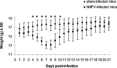
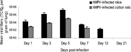
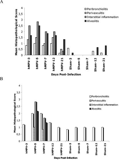
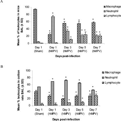
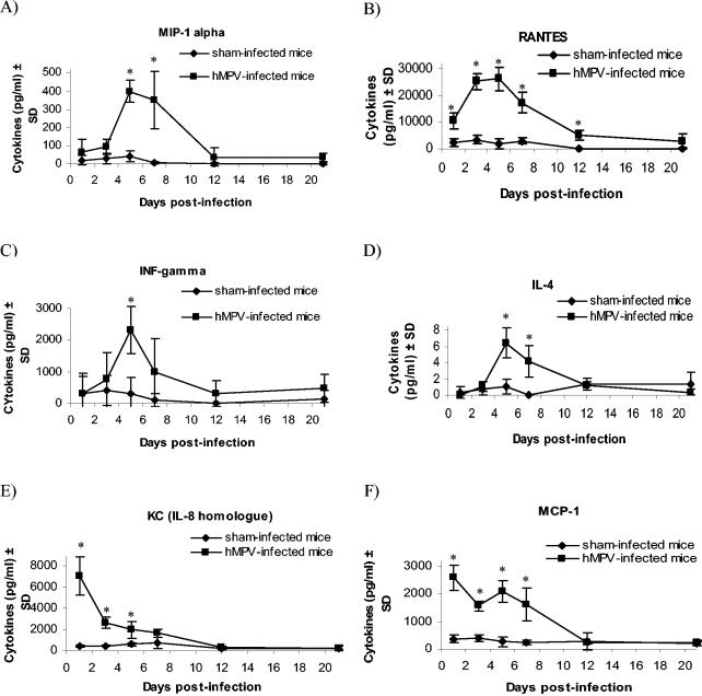
Human metapneumovirus (hMPV) is a newly described member of the Paramyxoviridae family causing acute respiratory tract infections, especially in young children. We studied the pathogenesis of this viral infection in two experimental small animal models (BALB/c mice and cotton rats). Significant viral replication in the lungs of both animals was found following an intranasal challenge of 10(8) 50% tissue culture infectious doses (TCID50) and persisted for <2 and <3 weeks in the case of cotton rats and mice, respectively. Peak viral loads were found on day 5 postinfection in both mice (mean of 1.92 x 10(7) TCID50/g lung) and cotton rats (mean of 1.03 x 10(5) TCID50/g). Clinical symptoms consisting of breathing difficulties, ruffled fur, and weight loss were noted in mice only around the time of peak viral replication. Most significant pulmonary inflammatory changes and peak expression of macrophage inflammatory protein 1alpha, gamma interferon, and RANTES occurred at the time of maximal viral replication (day 5) in both models. Cellular infiltration occurred predominantly around and within alveoli and persisted for at least 21 days in mice, whereas it was more limited in time with more peribronchiolitis in cotton rats. Both animal models would be of great value in evaluating different therapeutic agents, as well as vaccine candidates against hMPV.


Figures

FIG. 1.
FIG. 1.
Daily mean weights of hMPV- and sham-infected mice. Eighteen mice were sacrificed at serial times postinfection (days 1, 3, 5, 7, 12, and 21), beginning with 108 mice per group. *, statistically significant differences (P < 0.05) in weight were observed on days 4 to 10 postinfection, based on the Mann-Whitney rank sum test. SD, standard deviation.
FIG. 2.
FIG. 2.
Human metapneumovirus replication in lungs of BALB/c mice and cotton rats. Six mice (solid bars) and six cotton rats (hatched bars) were sacrificed on days 1, 3, 5, 7, 12, and 21 postinfection, and their lungs were removed. Lung homogenates were serially diluted and inoculated on LLC-MK2 cells for viral titration.
FIG. 3.
FIG. 3.
Histopathological scores in hMPV- and sham-infected mice and cotton rats. The degree of lung inflammation (mean histopathologic score) was evaluated for peribronchial, perivascular, interstitial, and alveolar areas in mice (A) and cotton rats (B). The histopathological score was defined as described previously (35).
FIG. 4.
FIG. 4.
Lung histopathology of hMPV- and sham-infected mice and cotton rats. (A) Six mice per group were sacrificed at different times postinfection, and their lungs were removed and fixed with 10% formalin. Thin sections of paraffin-embedded lung tissues were cut and stained with hematoxylin and eosin. A representative section (magnification, ×10) is shown for sham-infected mice on day 3 and hMPV-infected mice on days 3, 5, 7, 12, and 21 postinfection. Lung histopathology of sham-infected mice at different time points was similar to that shown on day 3. (B) Representative sections (magnification, ×20) of cotton rat lungs are shown for sham-infected rats on day 3 and for the same days postinfection as the mice.
FIG. 5.
FIG. 5.
Recruitment of inflammatory cells in BAL of hMPV- and sham-infected mice and cotton rats. (A) Six mice were sacrificed at different times postinfection (days 1, 3, 5, and 7), and BAL fluids were collected. Total leukocyte populations were determined using an hemacytometer, and then 2.5 × 10⁵ cells were spotted on a slide with a cytospin. Specific cell populations were distinguished with a hematoxylin-eosin stain. Leukocyte counts were similar on all days in sham-infected mice. No changes in the amount of eosinophils were observed. (B) Six cotton rats were sacrificed at different times postinfection (days 1, 3, 5, and 7), and BAL fluids were collected. A total of 104 cells were used to perform a fluorescence-activated cell sorter assay to distinguish specific cell populations. *, statistically significant differences (P < 0.05) were observed between hMPV-infected and sham-infected groups, based on the Mann-Whitney rank sum test. SD, standard deviation.
FIG. 6.
FIG. 6.
Cytokine/chemokine levels in lung homogenates of hMPV- and sham-infected mice. Six mice from both hMPV- and sham-infected groups were sacrificed on days 1, 3, 5, 7, 12, and 21 postinfection, and 50 μl of their lung homogenates was used to quantify MIP-1α (A), RANTES (B), IFN-γ (C), IL-4 (D), KC (E), and MCP-1 (F) by enzyme-linked immunosorbent assay. *, statistically significant differences (P < 0.05) were observed between hMPV- and sham-infected groups, based on Student's t test when data were normally distributed and on the Mann-Whitney rank sum test when data were not normally distributed. SD, standard deviation.
FIG. 7.
FIG. 7.
Cytokine/chemokine mRNA expression in lungs homogenates of hMPV- and sham-infected cotton rats. Cotton rat lung cytokines mRNA were quantified and normalized with β-actin. A reverse transcription reaction using oligo(dT) primers was done with total lung RNA, and 50 pg of cDNA constructs was loaded to run the PCR. Detection of lung cytokines by Southern blotting was semiquantitatively measured (+ to +++). Plus signs indicate the mean degree of intensity for six cotton rats for the bands targeting the specific cytokine.

References

    1. Alvarez, R., K. S. Harrod, W. J. Shieh, S. Zaki, and R. A. Tripp. 2004. Human metapneumovirus persists in BALB/c mice despite the presence of neutralizing antibodies. J. Virol. 78:14003-14011. - PMC - PubMed
    1. Arnold, R., F. Werner, B. Humbert, H. Werchau, and W. Konig. 1994. Effect of respiratory syncytial virus-antibody complexes on cytokine (IL-8, IL-6, TNF-alpha) release and respiratory burst in human granulocytes. Immunology 82:184-191. - PMC - PubMed
    1. Barends, M., L. G. de Rond, J. Dormans, M. van Oosten, A. Boelen, H. J. Neijens, A. D. Osterhaus, and T. G. Kimman. 2004. Respiratory syncytial virus, pneumonia virus of mice, and influenza A virus differently affect respiratory allergy in mice. Clin. Exp. Allergy 34:488-496. - PubMed
    1. Becker, S., J. Quay, and J. Soukup. 1991. Cytokine (tumor necrosis factor, IL-6, and IL-8) production by respiratory syncytial virus-infected human alveolar macrophages. J. Immunol. 147:4307-4312. - PubMed
    1. Biacchesi, S., M. H. Skiadopoulos, G. Boivin, C. T. Hanson, B. R. Murphy, P. L. Collins, and U. J. Buchholz. 2003. Genetic diversity between human metapneumovirus subgroups. Virology 315:1-9. - PubMed