Nairovirus RNA sequences expressed by a Semliki Forest virus replicon induce RNA interference in tick cells
- PMID: 15994788
- PMCID: PMC1168744
- DOI: 10.1128/JVI.79.14.8942-8947.2005
Nairovirus RNA sequences expressed by a Semliki Forest virus replicon induce RNA interference in tick cells
Abstract
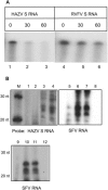
We report the successful infection of the cell line ISE6 derived from Ixodes scapularis tick embryos by the tick-borne Hazara virus (HAZV), a nairovirus in the family Bunyaviridae. Using a recombinant Semliki Forest alphavirus replicon that replicates in these cells, we were able to inhibit replication of HAZV, and we showed that this blockage is mediated by the replication of the Semliki Forest alphavirus replicon; the vector containing the HAZV nucleoprotein gene in sense or antisense orientation efficiently inhibited HAZV replication. Moreover, expression of a distantly related nucleoprotein gene from Crimean-Congo hemorrhagic fever nairovirus failed to induce HAZV silencing, indicating that the inhibition is sequence specific. The resistance of these cells to replicate HAZV correlated with the detection of specific RNase activity and 21- to 24-nucleotide-long small interfering RNAs. Altogether, these results strongly suggest that pathogen-derived resistance can be established in the tick cells via a mechanism of RNA interference.
Figures


References
-
- Adelman, Z. N., C. D. Blair, J. O. Carlson, B. J. Beaty, and K. E. Olson. 2001. Sindbis virus-induced silencing of dengue viruses in mosquitoes. Insect Mol. Biol. 10:265-273. - PubMed
-
- Adelman, Z. N., I. Sanchez-Vargas, E. A. Travanty, J. O. Carlson, B. J. Beaty, C. D. Blair, and K. E. Olson. 2002. RNA silencing of dengue virus type 2 replication in transformed C6/36 mosquito cells transcribing an inverted-repeat RNA derived from the virus genome. J. Virol. 76:12925-12933. - PMC - PubMed
-
- Aljamali, M. N., A. D. Bior, J. R. Sauer, and R. C. Essenberg. 2003. RNA interference in ticks: a study using histamine binding protein dsRNA in the female tick Amblyomma americanum. Insect Mol. Biol. 12:299-305. - PubMed
-
- Aljamali, M. N., J. R. Sauer, and R. C. Essenberg. 2002. RNA interference: applicability in tick research. Exp. Appl. Acarol. 28:89-96. - PubMed
-
- Begum, F., C. L. Wisseman, Jr., and J. Casals. 1970. Tick-borne viruses of West Pakistan. II. Hazara virus, a new agent isolated from Ixodes redikorzevi ticks from the Kaghan Valley, W. Pakistan. Am. J. Epidemiol. 92:192-194. - PubMed
Publication types
MeSH terms
Substances
LinkOut - more resources
Full Text Sources
Other Literature Sources

