. 2005 Jul;79(14):8979-90.
doi: 10.1128/JVI.79.14.8979-8990.2005.

The replicative fitness of primary human immunodeficiency virus type 1 (HIV-1) group M, HIV-1 group O, and HIV-2 isolates

Affiliations

The replicative fitness of primary human immunodeficiency virus type 1 (HIV-1) group M, HIV-1 group O, and HIV-2 isolates

Kevin K Ariën et al. J Virol. 2005 Jul.

Abstract

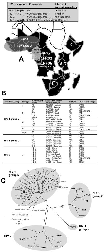
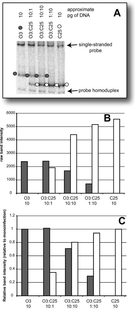
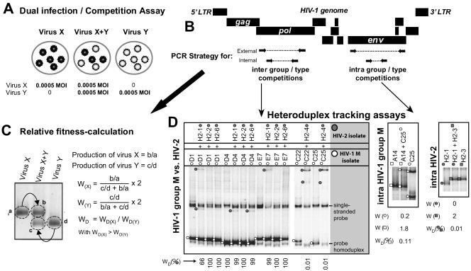
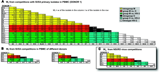
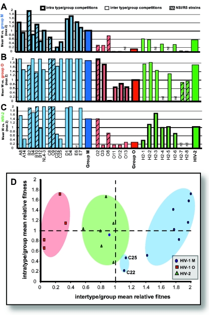
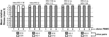
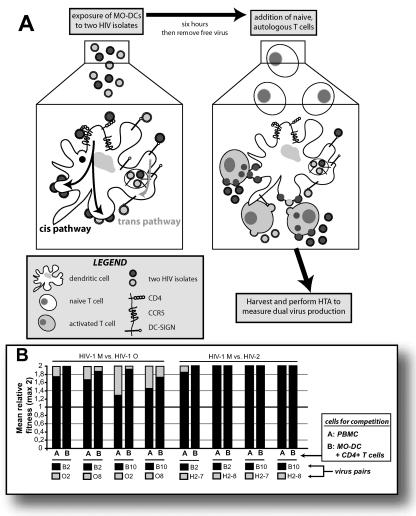
The main (M) group of human immunodeficiency virus type 1 (HIV-1) is responsible for the global AIDS epidemic while HIV-1 group O (outlier) and HIV type 2 are endemic only in west and central Africa. The failure of HIV-2 and especially HIV-1 group O to spread following the initial zoonotic jumps is not well understood. This study was designed to examine the relative replicative capacities between these human lentiviruses. A pairwise competition experiment was performed with peripheral blood mononuclear cells with eight HIV-2 isolates, 6 group O viruses, and 15 group M viruses of subtype A (2 viruses), B (5 viruses), C (4 viruses), D (2 viruses) and CRF01_AE (2 viruses). HIV-1 group M isolates of any subtype were typically 100-fold-more fit than group O or HIV-2 strains when competed in peripheral blood mononuclear cells from various humans. This order in replicative fitness was also observed when virus pairs were added to human dendritic cells and then cocultured with primary, quiescent T cells, which is the model for HIV-1 transmission. These results suggest that reduced replicative and transmission fitness may be contributing to the low prevalence and limited geographical spread of HIV-2 and group O HIV-1 in the human population.


Figures

FIG. 1.
FIG. 1.
(A) Prevalence of HIV in sub-saharan Africa is summarized in the grey panel. Sub-saharan Africa is further subdivided into various regions based on group M subtype prevalence. The size of the letters provides an indication of the prevalence of that subtype or CRF in a given region. For example, northeastern countries of Africa (i.e., Ethiopia, Eritriea, and Somalia) are dominated by subtype C. The grey shaded areas show the approximate range of HIV-2, HIV-1/HIV-2, and HIV-1 group O in Africa. (B) Virus characteristics of the isolates used in the dual infection/competition experiments. Most isolates have the syncytium-inducing phenotype (as determined with MT-2 cell line) and are able to use either the CXCR4 chemokine receptor exclusively as entry coreceptor or are dualtropic for both CXCR4 and CCR5 (as determined with a U87.CD4 cell line expressing either CXCR4 or CCR5). One isolate, O13, was initially identified as an SI isolate but was later found to be an NSI/R5 HIV-1 group O isolate through phenotypic analyses (see Materials and Methods). (C) Analysis of genotypic relationships of diverse HIV isolates by the neighbor-joining method. Phylogenetic neighbor-joining trees were constructed from alignments of the 400-nucleotide pol gene fragments from several HIV reference strains and 22 HIV isolates employed in the subsequent fitness analyses. Accession numbers for all pol gene fragments are provided in Materials and Methods. ** and *, bootstrap resampling values of 80 to 95% and >95%, respectively. Branch lengths are drawn to scale, and the scale bar represents 0.1 substitution per nucleotide.
FIG. 2.
FIG. 2.
Testing for preferential probe annealing in the heteroduplex tracking. The heteroduplex tracking assay is used to detect the PCR products originating from the two HIV isolates added to dual infections. The external PCR products from quantitation study described in Fig. S1 in the supplemental material were mixed for mock PCR amplication-HTA analyses. Several pairs of group M plus group O, group M plus HIV-2, and group O plus HIV-2 PCR products were mixed at 10:1, 10:10, and 1:10 ratios and then PCR amplified using the appropriate set of nested primers. Ten microliters of this amplified mixture was then added with 1 pmol of radiolabeled probe to an HTA reaction mixture (see Materials and Methods). The heteroduplexes from this mock competition and from mock monoinfections (10 pg of DNA added to the nested PCR and then annealed to the probe) were resolved on a 6% PAG and quantified on a Bio-Rad phosphorimager screen. (A) Image of a 6% PAG containing an HTA of the mock competition between O3 and C25 HIV-1 DNA, which was then probed with a group M HXB2 probe. (B) The raw intensity of eachheteroduplex band in this mock HTA was plotted. (C) The intensity of each heteroduplex band was then plotted relative to the heteroduplex of only the PCR amplified DNA from mock monoinfections. It is important to note that each lane of the HTA shown in panel A contains the same amount of PCR-amplified DNA. The HTA shown in panel A and the analyses shown in panels B and C were repeated with the same samples using the group O isolate ESP1 probe. Similar mock analyses were performed on pairwise mock competitions involving HTAs, following nested amplification of the D1, C25, O2, O3, O11, H2-2, and H2-3 templates (data not shown).
FIG. 3.
FIG. 3.
Schematic representation and sample results of HIV competition experiments, heteroduplex tracking assays, and fitness analyses. (A) Virus was added alone or in pairs to PHA-stimulated and IL-2-treated PBMCs at an equal MOI of 0.0005. Cells were washed after 24 h to remove residual virus. Cells and virus supernatant were harvested at day 10 and lysed for subsequent DNA extraction. (B) Extracted DNA from dual infections was PCR amplified using env primer pairs specific for HIV-1 M, HIV-1 O, and HIV-2 (see Table S1 in the supplemental material). RT primers were also designed to amplify DNA from HIV-1 M/O, HIV-1 O/HIV-2, and HIV-1 M/HIV-2 dual infections (see Table S1 in the supplemental material). (C) Relative fitness values (W) and a fitness ratio referred to as fitness difference (WD) were derived as shown. (D) Samples of HTAs and fitness calculations for HIV-1 group M versus HIV-2 competitions, as well as intragroup M and intratype HIV-2 competitions. Radiolabeled, PCR-amplified DNA from viruses A8, D1, and H2-2 were used as probes.
FIG. 4.
FIG. 4.
Fitness difference values from all of the dual virus competitions involving 28 HIV isolates of type 1 group M and group O and type 2. (A) Matrix showing relative fitness differences (WD) of pairwise dual infection competitions in PHA/IL-2-stimulated PBMCs from a healthy HIV-negative blood donor. Relative fitness values can be derived from the equation x = 2y/(y + 1), where y = fitness difference (WD), x = fitness (Wx) of the numerator, and 2 − x = fitness (W2−x) of the denominator in the equation WD = Wx/W2−x. (B) Matrices, showing relative fitness differences (WD) of two subset pairwise competitions performed with PHA/IL-2-stimulated PBMCs from two additional HIV negative blood donors (one Caucasian and one Bantu African). Finally, eight NSI/R5 primary isolates (two of group M subtype B, two of group M subtype C, two of group O, and two of HIV-2) were also used for a pairwise competition with PHA/IL-2-treated PBMCs (C). It should be noted that due to limited quantities of virus stocks, some competitions in the complete pairwise matrix could not be performed.
FIG. 5.
FIG. 5.
(A to C) The mean relative fitness values (maximum, 2) for intratype/intragroup and intertype/intergroup competitions in PBMC involving both SI and NSI HIV-1 group M and group O and HIV-2 primary isolates. (D) Intertype/intergroup versus intratype/intragroup mean relative fitness of all SI isolates. Fitness differences were derived from competitions involving HIV-1 M and O and HIV-2 are shown in Fig. 4.
FIG. 6.
FIG. 6.
Comparing the relative fitness of various primary HIV isolates in a subset of competitions performed with PBMCs of three different HIV-negative blood donors (two Caucasians and one Bantu African). Mean relative fitness values (maximum, 2) are shown for two intragroup/intratype and three intergroup/intertype competitions in the three different blood donors. A full set of relative fitness values is shown in Fig. 4B.
FIG. 7.
FIG. 7.
(A) Schematic representation of the HIV dual infection of the MO-DC-CD4+ T-cell cultures. MO-DCs were first exposed to a pair of HIV isolates for 6 h, after which free virus was washed away and CD4+ T cells were added. The prevailing theory suggests that MO-DC can mediate T-cell stimulation and transfer of virus. CD4+ T cells are either infected through trans exposure from virus produced from infected MO-DCs or presented in cis via DC-SIGN-mediated endocytosis and release. (B) Fitness difference values derived from pairwise competitions with NSI/R5 HIV-1 M and O and HIV-2 primary isolates in PBMCs and MO-DCs plus CD4+ T-cell cocultures.

Similar articles

Cited by

References

    1. Anderson, R. M., and R. M. May. 1996. The population biology of the interaction between HIV-1 and HIV-2: coexistence or competitive exclusion? AIDS 10:1663-1673. - PubMed
    1. Anderson, R. M., and R. M. May. 1991. Infectious diseases of humans. Oxford University Press, Oxford, United Kingdom.
    1. Arts, E. J., and M. E. Quinones-Mateu. 2003. Sorting out the complexities of HIV-1 fitness. AIDS 17:780-781. - PubMed
    1. Ayouba, A., P. Mauclere, P. M. Martin, P. Cunin, J. Mfoupouendoun, B. Njinku, S. Souquieres, and F. Simon. 2001. HIV-1 group O infection in Cameroon, 1986 to 1998. Emerg. Infect. Dis. 7:466-467. - PMC - PubMed
    1. Ball, S. C., A. Abraha, K. R. Collins, A. J. Marozsan, H. Baird, M. E. Quiñones-Mateu, A. Penn-Nicholson, M. Murray, N. Richard, M. Lobritz, P. A. Zimmerman, T. Kawamura, A. Blauvelt, and E. J. Arts. 2003. Comparing the ex vivo fitness of CCR5-tropic human immunodeficiency virus type 1 isolates of subtypes B and C. J. Virol. 77:1021-1038. - PMC - PubMed

Publication types

MeSH terms