. 2005 Jul;79(14):9134-44.
doi: 10.1128/JVI.79.14.9134-9144.2005.

Analysis of human immunodeficiency virus type 1 Gag ubiquitination

Affiliations

Analysis of human immunodeficiency virus type 1 Gag ubiquitination

Eva Gottwein et al. J Virol. 2005 Jul.

Abstract

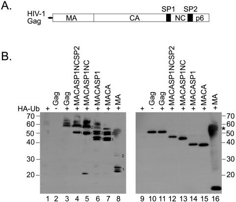
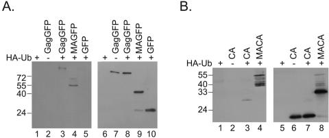
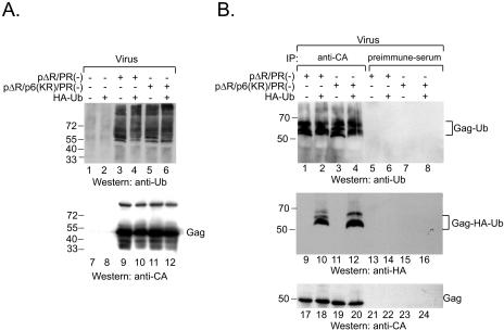
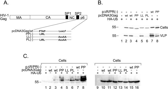
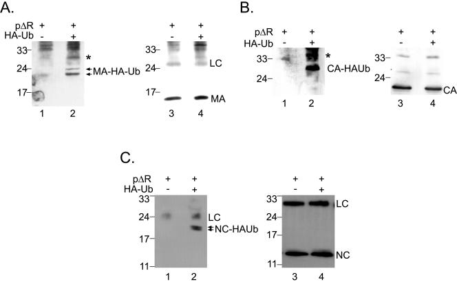
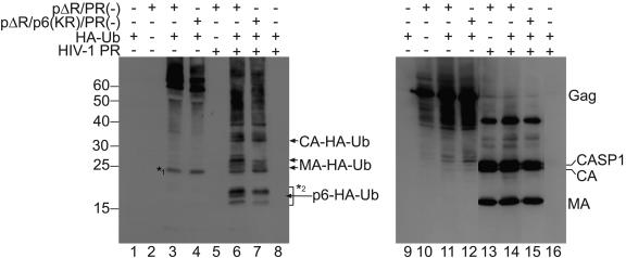
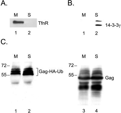
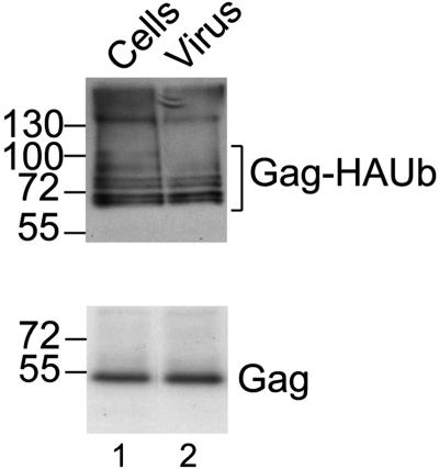
Ubiquitin is important for the release of human immunodeficiency virus type 1 (HIV-1) and several other retroviruses, but the functional significance of Gag ubiquitination is unknown. To address this problem, we decided to analyze Gag ubiquitination in detail. A low percentage of the HIV-1 p6 protein has previously been shown to be ubiquitinated, and published mutagenesis data suggested that Gag ubiquitination is largely lost upon mutation of the two lysine residues in p6. In this study, we show that Gag proteins lacking the p6 domain or the two lysine residues within p6 are ubiquitinated at levels comparable to those of the wild-type Gag protein. We detected monoubiquitinated forms of the matrix (MA), capsid (CA), and nucleocapsid (NC) proteins in mature virus preparations. Protease digestion of Gag polyproteins extracted from immature virions indicated that ubiquitinated MA, CA, and possibly NC are as abundant as ubiquitinated p6. The HIV-1 late-domain motifs PTAP and LRSLF were not required for Gag ubiquitination, and mutation of the PTAP motif even resulted in an increase in the amount of Gag-Ub conjugates detected. Finally, at steady state, ubiquitinated Gag proteins were not enriched in either membrane-associated or virus-derived Gag fractions. In summary, these results indicate that HIV-1 Gag can be monoubiquitinated in all domains and that ubiquitination of lysine residues outside p6 may thus contribute to viral release and/or infectivity.


Figures

FIG. 1.
FIG. 1.
(A) Schematic representation of the HIV-1 Gag polyprotein. (B) Ubiquitination of C-terminally truncated Gag proteins. Truncated Gag proteins were expressed from pcDNA3-based Rev-independent Gag expression constructs. Proteins are designated by the domains that are present in the protein (panel A). The indicated proteins were expressed in the presence (+) or absence (−) of HA-Ub in 293T cells and, 48 h after transfection, immunoprecipitated with sheep anti-CA antiserum (lanes 1 to 7) or rabbit anti-MA antiserum (lane 8). The immunoprecipitates were examined by Western blotting with an anti-HA antibody to visualize Gag-HA-Ub conjugates (left). Proteins that are presumably modified by one or two ubiquitin moieties are marked 1 and 2, respectively. To visualize the recovered Gag proteins, the blot was reprobed with rabbit anti-MA antibodies (right). Since MA was immunoprecipitated with the same antibody, the heavy chain was also detected on the blot in this case (lane 16, marked by an asterisk). Molecular mass standards (in kDa) are shown at the side of each panel.
FIG. 2.
FIG. 2.
(A) Ubiquitination of MA-GFP. Gag-GFP, MA-GFP, or GFP alone was expressed in the presence (+) or absence (−) of HA-Ub as indicated. Cell lysates were immunoprecipitated with rabbit anti-GFP antiserum and probed for HA-Ub modification of precipitated proteins by Western blotting with anti-HA antibodies (left). The blot was reprobed using a monoclonal anti-GFP antibody (right). Molecular mass standards (in kDa) are shown on the left side. (B) Ubiquitination of CA. CA or MACA was expressed from pcDNA3-based, Rev-independent expression constructs in the presence (+) or absence (−) of HA-Ub as indicated. Immunoprecipitation was performed using rabbit anti-CA antiserum. HA-Ub-modified proteins were detected by Western blotting with an anti-HA antibody (left). The blot was reprobed with anti-CA antibodies (right). Molecular mass standards (in kDa) are shown on the left.
FIG. 3.
FIG. 3.
Comparison of the expression levels of viral proteins from pNL4-3 and pΔR. 293T cells were transfected with pcDNA3 (left lane in each blot), pNL4-3 (middle lane), or pΔR (right lane). Virions were pelleted through sucrose cushions and examined by Western blotting using anti-CA (top left), anti-MA (top middle), anti-NC (top right), anti-gp41 (lower left), or anti-RT (lower right) antibodies. The variant RT proteins expressed from pΔR travel slightly faster than the wild-type RT proteins (compare lane 15 to lane 14). HIV-1-specific proteins are identified on the right side of each panel.
FIG. 4.
FIG. 4.
HIV-1 Gag lacking lysine residues in p6 is ubiquitinated. (A) The wild type (lanes 3, 4, 9, and 10) or p6 lysine mutant (lanes 5, 6, 11, and 12) pΔR/PR(−) was cotransfected with pHA-Ub as indicated (+). Virions were pelleted through sucrose cushions and directly analyzed by SDS-PAGE and Western blotting with an anti-Ub antibody (top). The blot was reprobed with anti-CA antibody (bottom). Molecular mass standards (in kDa) are shown at the side of each panel. (B) Transfection and virion preparation were carried out as described for panel A, but the virus pellets were resuspended in a buffer containing 1% SDS and boiled for 10 min. Subsequently, the buffer was adjusted to RIPA buffer and samples were split into two aliquots, one of which was immunoprecipitated (IP) with anti-CA antiserum (lanes 1 to 4, 9 to 12, and 17 to 20) while the other was immunoprecipitated with preimmune serum (lanes 5 to 8, 13 to 16, and 21 to 24). Gag samples were adjusted to equal amounts of Gag (as quantified by p24 ELISA), and the whole precipitates were analyzed in the case of the negative controls. Immunoprecipitates were resolved by SDS-PAGE and analyzed by Western blotting with anti-Ub antibodies (top). The membrane was stripped and consecutively reprobed with anti-HA (middle) and anti-CA (bottom) antibodies.
FIG. 5.
FIG. 5.
(A) Schematic representation of the PTAP and LRSLF mutant Gag proteins used. (B) 293T cells were cotransfected using (+) the indicated plasmids. Twenty hours after transfection, cells (top) and virions (bottom) were harvested and analyzed by Western blotting with an anti-CA antibody. Molecular mass standards (in kDa) are shown at the side of each panel. (C) The remaining cell extracts were immunoprecipitated with anti-CA antiserum and analyzed by SDS-PAGE and Western blotting with an anti-HA antibody (left). The blot was reprobed with an anti-CA antibody (right). Molecular mass standards (in kDa) are shown on the left side.
FIG. 6.
FIG. 6.
Ubiquitination of virus-derived HIV-1 Gag proteins. 293T cells were cotransfected with pΔR and pcDNA3 (lanes 1 and 3) or pΔR and an HA-Ub expression plasmid (lanes 2 and 4). Forty-eight hours after transfection, virions were pelleted through sucrose cushions, lysed in RIPA buffer, and immunoprecipitated with antiserum against MA (A), CA (B), or NC (C). Immunoprecipitates were resolved by SDS-PAGE and subjected to Western blotting with anti-HA antibodies (left) or HIV-1-specific antibodies (right). HA-Ub-modified proteins are identified at the sides of the left panels. Molecular mass standards (in kDa) are shown on the left side of each panel. Bands marked by an asterisk probably represent diubiquitinated versions of the respective HIV-1 proteins. LC indicates detection of the light chain of the antibody used for immunoprecipitation.
FIG. 7.
FIG. 7.
Digestion of ubiquitinated Gag by exogenously added HIV-1 PR. 293T cells were transfected with either pHA-Ub alone or pΔR/PR(−) alone or cotransfected with pHA-Ub and pΔR/PR(−) or pΔR/p6(KR)/PR(−) as indicated. Forty-eight hours after transfection, virions were pelleted through sucrose cushions and resuspended in PR cleavage buffer. The virions were lysed by adjusting the buffer to 0.1% Triton and divided into two aliquots. While one aliquot was left untreated (lanes 1 to 4 and 9 to 12), the second aliquot was treated with 120 nM HIV-1 PR for 1 hour (lanes 5 to 8 and 13 to 16). After protease digestion, samples were analyzed by SDS-PAGE and Western blotting with anti-HA antibodies (left). The blot was reprobed with a mixture of anti-CA and anti-MA antisera to confirm Gag cleavage. The band marked by *1 represents a form of Ub that is detected in the absence of Gag cleavage. The bands marked by *2 represent ubiquitinated Gag cleavage products other than p6, MA, and CA, possibly NC or SP2.
FIG. 8.
FIG. 8.
Ubiquitinated Gag is not enriched in membrane fractions. 293T cells were cotransfected with pHA-Ub and pΔR/PR(−) and, 48 h after transfection, subjected to membrane flotation. The visible membrane (M) and soluble (S) fractions were collected. A small part of each fraction was analyzed by Western blotting for the fractionation markers TfnR (A) and 14-3-3γ (B). The remaining part of each fraction was immunoprecipitated for the CA protein and analyzed by Western blotting using anti-HA antibodies (C, lanes 1 and 2). The blot was reprobed with monoclonal anti-CA antibodies (C, lanes 3 and 4) to confirm that similar amounts of Gag were analyzed.
FIG. 9.
FIG. 9.
Ubiquitinated Gag is not enriched in the virus. 293T cells were cotransfected with pHA-Ub and ΔR/PR(−). Forty-eight hours after transfection, virions were pelleted through sucrose cushions and cells were detached from the culture dish and washed with PBS. Virus and cell pellets were boiled in a buffer containing 1% SDS for 10 min. The cell and virus lysates were adjusted to RIPA buffer, cleared, and immunoprecipitated with anti-CA antiserum. Samples were normalized for equal CA contents (by Western blotting with anti-CA antibody) (bottom) and analyzed by Western blotting with an anti-HA antibody (top).

Similar articles

Cited by

References

    1. Abacioglu, Y. H., T. R. Fouts, J. D. Laman, E. Claassen, S. H. Pincus, J. P. Moore, C. A. Roby, R. Kamin-Lewis, and G. K. Lewis. 1994. Epitope mapping and topology of baculovirus-expressed HIV-1 gp160 determined with a panel of murine monoclonal antibodies. AIDS Res. Hum. Retrovir. 10:371-381. - PubMed
    1. Adachi, A., H. E. Gendelman, S. Koenig, T. Folks, R. Willey, A. Rabson, and M. A. Martin. 1986. Production of acquired immunodeficiency syndrome-associated retrovirus in human and nonhuman cells transfected with an infectious molecular clone. J. Virol. 59:284-291. - PMC - PubMed
    1. Bohne, J., and H. G. Krausslich. 2004. Mutation of the major 5′ splice site renders a CMV-driven HIV-1 proviral clone Tat-dependent: connections between transcription and splicing. FEBS Lett. 563:113-118. - PubMed
    1. Bouamr, F., J. A. Melillo, M. Q. Wang, K. Nagashima, M. de Los Santos, A. Rein, and S. P. Goff. 2003. PPPYVEPTAP motif is the late domain of human T-cell leukemia virus type 1 Gag and mediates its functional interaction with cellular proteins Nedd4 and Tsg101. J. Virol. 77:11882-11895. - PMC - PubMed
    1. Freed, E. O. 1998. HIV-1 gag proteins: diverse functions in the virus life cycle. Virology 251:1-15. - PubMed

Publication types