. 2005 Jul;79(14):9157-67.
doi: 10.1128/JVI.79.14.9157-9167.2005.

Genetic variability and molecular evolution of the human respiratory syncytial virus subgroup B attachment G protein

Affiliations

Genetic variability and molecular evolution of the human respiratory syncytial virus subgroup B attachment G protein

Kalina T Zlateva et al. J Virol. 2005 Jul.

Abstract

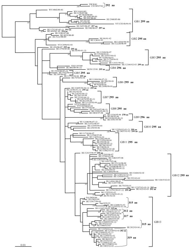
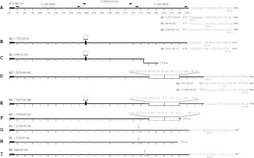
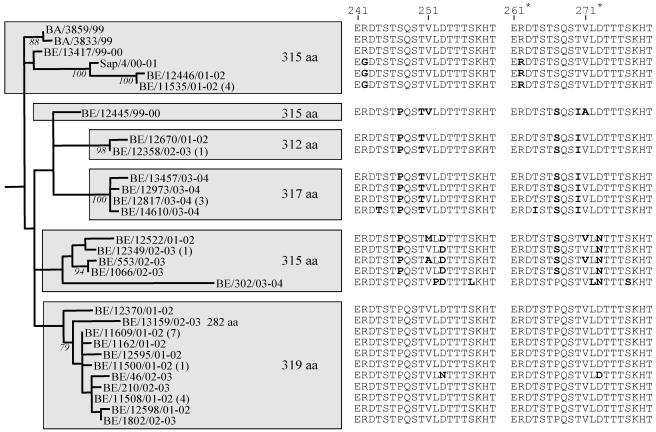
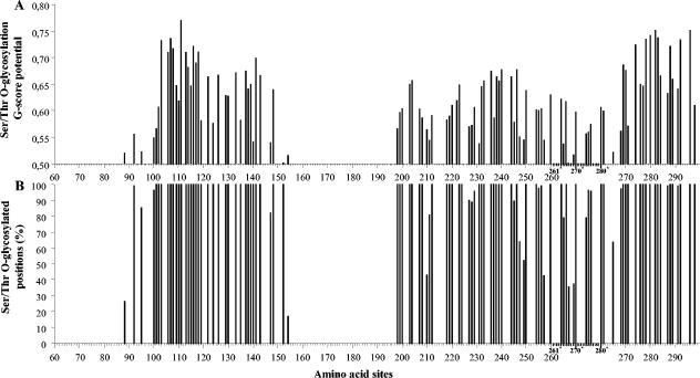
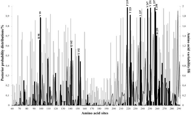
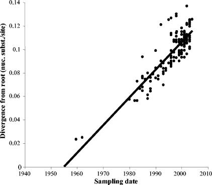
Human respiratory syncytial virus (HRSV) is the most important cause of acute respiratory disease in infants. Two major subgroups (A and B) have been identified based on antigenic differences in the attachment G protein. Antigenic variation between and within the subgroups may contribute to reinfections with these viruses by evading the host immune responses. To investigate the circulation patterns and mechanisms by which HRSV-B viruses evolve, we analyzed the G protein genetic variability of subgroup B sequences isolated over a 45-year period, including 196 Belgian strains obtained over 22 epidemic seasons (1982 to 2004). Our study revealed that the HRSV-B evolutionary rate (1.95 x 10(-3) nucleotide substitutions/site/year) is similar to that previously estimated for HRSV-A (1.83 x 10(-3) nucleotide substitutions/site/year). However, natural HRSV-B isolates appear to accommodate more drastic changes in their attachment G proteins. The most recent common ancestor of the currently circulating subgroup B strains was estimated to date back to around the year 1949. The divergence between the two major subgroups was calculated to have occurred approximately 350 years ago. Furthermore, we have identified 12 positively selected sites in the G protein ectodomain, suggesting that immune-driven selective pressure operates in certain codon positions. HRSV-A and -B strains have similar phylodynamic patterns: both subgroups are characterized by global spatiotemporal strain dynamics, where the high infectiousness of HRSV permits the rapid geographic spread of novel strain variants.


Figures

FIG.1.
FIG.1.
HRSV-B G protein maximum likelihood phylogenetic tree. The nucleotide sequences of the G protein gene of the Belgian isolates (BE) were compared with those from Buenos Aires (BA), District of Columbia (CH), Montevideo (MON), New Mexico (NM), New York (NY), Sweden (SW), and West Virginia (WV). The reference strain SW/8/60 was used as outgroup sequence in the tree. Numbers at internal nodes represent bootstrap percentages, as determined for 1,000 iterations by the neighbor-joining method. Only bootstrap values greater than 75% are shown. The italicized numbers in brackets at the terminal nodes correspond to the number of identical sequences. The genetic clusters obtained in the analysis are indicated by the square brackets and assigned as GB1 to GB13. The predicted G protein lengths are designated in boldface; exceptions are indicated next to the corresponding sequence.
FIG. 2.
FIG. 2.
Schematic diagrams presenting the predicted G protein length variability among the HRSV-B Belgian isolates. The linear presentations are given according to the reference strain WV/B1/85 with the two variable regions and the central conserved region (residues 153 to 221) indicated above the top diagram. The absolutely conserved amino acid region (amino acid positions 164 to 187) among the HRSV-B isolates is indicated with a double line (A). The underlined sequences present the three alternative termination codons described previously (32), and the predicted full protein lengths of Belgian representative sequences are given. The six-nucleotide deletion is indicated with a filled box (B, C, and E), the 60-nt duplication is indicated with a large open and divided box (D to F), and the three-nucleotide insertions are indicated with a small open box (G to I). The position of the frame-shifting in the frameshift mutant is indicated (•) in panel C. The two premature terminated sequences with their respective protein lengths are presented in panels F and H.
FIG. 3.
FIG. 3.
Phylogenetic tree of the GB13 lineage containing HRSV-B strains with the 60-nt duplicated region isolated from Belgium (BE), Buenos Aires (BA), and Sapporo (Sap). Bootstrap values above 75% for 1,000 iterations are shown at the branch. The amino acid sequence of the duplicated region is shown next to each isolate with nonsynonymous substitutions presented in boldface.
FIG. 4.
FIG. 4.
Predicted Ser/Thr O-glycosylated residues in the HRSV-B viruses presented according to reference strain WV/B1/85. (A) Best general score potentials (G score) of the predicted O-glycosylated Ser/Thr sites. (B) Percentages of the HRSV-B sequences in which a particular Ser/Thr is predicted to be O glycosylated. The numbers with a superscript asterisk and the scale lines in bold indicate the amino acid positions in the duplicated 60-nt region identified in some HRSV-B strains.
FIG. 5.
FIG. 5.
Posterior probabilities of sites for different classes along the G protein ectodomain region under the discrete model M3. This model assumes three classes of sites in the gene: positive sites (black bars), neutral sites (open graph), and negative sites (gray bars). Positively selected amino acid sites with posterior probabilities above 50% are indicated according to strain WV/B1/85. Amino acid variability, measured by entropy (Hi), is plotted to the second y axis.
FIG. 6.
FIG. 6.
Linear root-to-tip regression plot presenting the correlation between the branch lengths and the sampling dates of the HRSV-B isolates included in this study.

References

    1. Anderson, L. J., J. C. Hierholzer, C. Tsou, R. M. Hendry, B. F. Fernie, Y. Stone, and K. McIntosh. 1985. Antigenic characterization of respiratory syncytial virus strains with monoclonal antibodies. J. Infect. Dis. 151:626-633. - PubMed
    1. Cane, P. A., and C. R. Pringle. 1995. Evolution of subgroup A respiratory syncytial virus: evidence for progressive accumulation of amino acid changes in the attachment protein. J. Virol. 69:2918-2925. - PMC - PubMed
    1. Cane, P. A., D. A. Matthews, and C. R. Pringle. 1993. Frequent polymerase errors observed in a restricted area of clones derived from the attachment (G) protein gene of respiratory syncytial virus. J. Virol. 67:1090-1093. - PMC - PubMed
    1. Cane, P. A., D. A. Matthews, and C. R. Pringle. 1991. Identification of variable domains of the attachment (G) protein of subgroup A respiratory syncytial viruses. J. Gen. Virol. 72:2091-2096. - PubMed
    1. Coggins, W. B., E. J. Lefkowitz, and W. M. Sullender. 1998. Genetic variability among group A and group B respiratory syncytial viruses in a children's hospital. J. Clin. Microbiol. 36:3552-3557. - PMC - PubMed

Publication types

MeSH terms

Substances

LinkOut - more resources