. 2005 Jul;79(14):9168-79.
doi: 10.1128/JVI.79.14.9168-9179.2005.

Suppressors of a host range mutation in the rabbitpox virus serpin SPI-1 map to proteins essential for viral DNA replication

Affiliations

Suppressors of a host range mutation in the rabbitpox virus serpin SPI-1 map to proteins essential for viral DNA replication

Benjamin G Luttge et al. J Virol. 2005 Jul.

Abstract

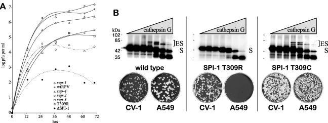
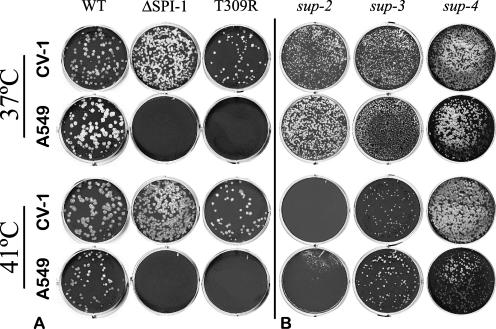
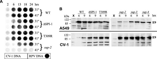
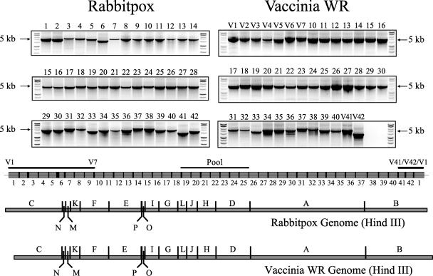
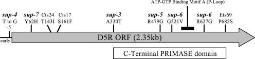
The orthopoxvirus serpin SPI-1 is an intracellular serine protease inhibitor that is active against cathepsin G in vitro. Rabbitpox virus (RPV) mutants with deletions of the SPI-1 gene grow on monkey kidney cells (CV-1) but do not plaque on normally permissive human lung carcinoma cells (A549). This reduced-host-range (hr) phenotype suggests that SPI-1 may interact with cellular and/or other viral proteins. We devised a genetic screen for suppressors of SPI-1 hr mutations by first introducing a mutation into SPI-1 (T309R) at residue P14 of the serpin reactive center loop. The SPI-1 T309R serpin is inactive as a protease inhibitor in vitro. Introduction of the mutation into RPV leads to the same restricted hr phenotype as deletion of the SPI-1 gene. Second-site suppressors were selected by restoration of growth of the RPV SPI-1 T309R hr mutant on A549 cells. Both intragenic and extragenic suppressors of the T309R mutation were identified. One novel intragenic suppressor mutation, T309C, restored protease inhibition by SPI-1 in vitro. Extragenic suppressor mutations were mapped by a new procedure utilizing overlapping PCR products encompassing the entire genome in conjunction with marker rescue. One suppressor mutation, which also rendered the virus temperature sensitive for growth, mapped to the DNA polymerase gene (E9L). Several other suppressors mapped to gene D5R, an NTPase required for DNA replication. These results unexpectedly suggest that the host range function of SPI-1 may be associated with viral DNA replication by an as yet unknown mechanism.


Figures

FIG. 1.
FIG. 1.
RPV host range can be restored by suppression of the SPI-1 T309R mutation. (A) Growth and spread of RPV mutants on A549 cells. Monolayers of A549 cells in six-well dishes were infected with ∼30 PFU of each RPV mutant under liquid medium at 37°C. Samples were harvested at 12-h intervals over 72 h, and the titers of the virus were determined on CV-1 cells. The titer of each virus with respect to time is shown. The initial concentration of virus was approximately 10 PFU per ml. RPVΔSPI-1 has a deletion of the SPI-1 ORF. T309R is the parental RPV SPI-1 P14 hr mutant. The RPV sup-1, sup-2, sup-3, and sup-4 mutants each contain suppressors of the SPI-1 T309R mutation. All suppressor isolates were selected on A549 cells by restored host range. Trend lines were drawn for each series as a fourth-order polynomial (Excel, Microsoft). Dotted trend lines indicate viruses which do not form plaques on A549 cells. While there is clearly growth of the T309R mutant in A549 cells relative to RPVΔSPI-1, the virus does not plaque on these cells. (B) Characterization of the SPI-1 T309C intragenic suppressor. Electrophoretic mobility shift assays with labeled SPI-1 protein (top panel) and the corresponding RPV host range phenotype in plaque assays (bottom panel) are shown. Wild-type SPI-1, SPI-1 T309R, or SPI-1 T309C DNA was cloned into the pTM1 vector for in vitro transcription and translation (TNT) to generate 35S-labeled SPI-1 protein. Increasing amounts of purified human cathepsin G (serine protease) were incubated at 37°C for 90 min with a constant amount of the labeled SPI-1 TNT product. The mixture was then resolved by 10% SDS-PAGE. Wild-type SPI-1 protein (∼45 kDa) generates two isoforms of a stable 1:1 complex with cathepsin G (∼25 kDa), where ES is enzyme-substrate. Formation of this complex is consistent with protease inhibitory function by a competitive and partially irreversible mechanism common to all inhibitory serpins. RPV containing the wild-type SPI-1 gene forms plaques on both CV-1 and A549 cells. The SPI-1 T309R mutation (middle) affects protease inhibition in vitro and leads to a reduced host range of the virus. Spontaneous reversion of RPV SPI-1 T309R to T309C (right) was isolated by genetic selection on A549 cells to restore plaque-forming ability. SPI-1 T309C protein shows a restored ability to form inhibitory complexes with cathepsin G in vitro that is comparable with wild-type SPI-1 protein.
FIG. 2.
FIG. 2.
Temperature sensitivity of RPV extragenic suppressor mutants. Equivalent dilutions of each RPV isolate were plaqued on monolayers of CV-1 and A549 cells under agarose media in six-well plates at either 37°C or 41°C to assay for temperature sensitivity (ts). (A) Deletion of the SPI-1 gene from RPVΔSPI-1 or inactivation of the SPI-1 gene by the T309R mutation reduces RPV host range on A549 cells but does not affect its ability to form plaques at 41°C on permissive cells (CV-1). (B) Extragenic suppressor mutations sup-2, sup-3, and sup-4 all restore plaque formation to the RPV SPI-1 T309R mutant on A549 cells. The RPV sup-2 mutant is temperature sensitive (ts) for growth. The RPV sup-3 mutant has a slightly reduced ability to form plaques at 41°C. The RPV sup-4 mutant exhibits no temperature sensitivity.
FIG. 3.
FIG. 3.
Mapping of the sup-2 mutation by marker rescue. The ts property of the RPV sup-2 mutant was used to map the sup-2 mutation by marker rescue. (A and B) Monolayers of CV-1 cells in six-well plates were infected with the RPV sup-2 mutant at an MOI of 0.003 PFU per cell (10,000 PFU per well). Cells were then transfected with individual wild-type virus DNA fragments from either the vaccinia virus (A) or RPV (B) genome, and recombinant viruses were identified by restored plaque formation (rescue) at 41°C. (A) Using a cosmid library of the wild-type vaccinia virus (WR) genome, only fragments pWR27-62 and pWR45-83 rescued RPV sup-2 mutant infections at 41°C (data not shown), which mapped the ts mutation to within 10 kb (indicated by dotted lines). (B) RPV sup-2 mutant- and RPV SPI-1 T309R-infected CV-1 cells incubated at 41°C in the absence of transfected DNA are shown for comparison (top). To refine the marker rescue analysis, DNA from the SPI-1 gene and the complete open reading frames E8R, E9L, and E10R (contiguous ORFs shown below) was amplified by PCR from wild-type RPV genomic DNA. Transfection of RPV sup-2 mutant-infected CV-1 cells with wild-type E9L DNA (center) rescues plaque formation at 41°C, which was not seen in any other transfection. As a negative control, mutant E9L PCR product amplified from RPV sup-2 mutant genomic DNA (sup-2 E9L, middle left) does not rescue the infection. Failure to rescue the RPV sup-2 mutant at 41°C upon transfection with wt SPI-1 DNA (middle right) also indicates that the ts phenotype conferred by sup-2 is independent of the parental SPI-1 mutation. A mutation within the E9L gene of the RPV sup-2 mutant, E9ΔH142, was later identified by DNA sequencing. This mutation (sup-2, bottom right) is at the junction of E9L and a hypothetical ORF named E ORF E. (C) Amino acid alignment of vertebrate poxvirus DNA polymerases from various genera illustrates that codon His142 (*) of RPV E9L is within a highly conserved C(Y/F)HC motif (shown in bold). RPV, rabbitpox (orthopox); MYX, myxoma (leporipox); SPV, swinepox (suipox); YLDV, Yaba-like disease virus (yatapox); ORF, orf virus (parapox); MCV, molluscum contagiosum virus (molluscipox); FPV, fowlpox (avipox). The start of a proposed 3′-5′ exonuclease domain (exo I) within the DNA polymerase protein is also indicated for reference (30, 47).
FIG. 4.
FIG. 4.
RPV host range mutant suppression by sup-2 does not depend on the hypothetical E ORF E gene. (A) Rescue of the RPV sup-2 mutant at 41°C in CV-1 cells by transfection with wt E9L PCR product replaces the E9ΔH142 allele in the RPV sup-2 mutant with wild-type E9L sequence (sup-2 wtE9). This recombinant virus does not grow in A549 cells, because it is a reconstruction of the parental RPV SPI-1 T309R mutant. Similarly, the RPV sup-2 mutant was later reconstructed from the RPV SPI-1 T309R mutant by replacing its wt E9L allele with mutant E9ΔH142 DNA using a variation on the marker rescue technique based on host range phenotype (hr) in A549 cells (data not shown). (B) DNA sequence analysis revealed that the sup-2 mutation also deletes the initiating Met1 codon from a hypothetical ORF designated E ORF E, which is embedded in the opposite orientation within the E9L ORF. To confirm that removal of the E ORF E gene by the sup-2 mutation has no effect on RPV host range, the E9ΔH142 allele of the RPV sup-2 mutant was replaced with wtE9ΔEORFE DNA by ts marker rescue. This removes E ORF E but restores the wt E9L gene. The phenotype of this recombinant (sup-2, wtE9ΔEORFE) is identical to the parental RPV SPI-1 T309R mutant.
FIG. 5.
FIG. 5.
Characterization of the RPV sup-2 mutant. (A) RPV DNA synthesis assay. CV-1 cells were infected with wild-type RPV or each designated mutant at an MOI of 10 PFU per cell at 31°C or 41°C. Samples were harvested at intervals up to 24 h postinfection, transferred to a nylon membrane by dot blot, and probed with digoxigenin-labeled RPV DNA to detect viral DNA synthesis by chemiluminescence. The CV-1 DNA and RPV DNA in the boxes contain control DNA samples for testing the hybridization specificity of the probe. (B) Western blot analysis of the E9L protein levels in infected cells. RPV-infected A549 (top) and CV-1 (bottom) cells were harvested at 6 and 9 h postinfection at 37°C. Steady-state levels of RPV DNA polymerase (∼116 kDa) were evaluated by Western blot analysis using a rabbit anti-VV-WR DNA polymerase antibody. X, uninfected cells.
FIG. 6.
FIG. 6.
PCR-generated libraries of RPV and VV-WR. Virtually the entire genomes of rabbitpox (RPV) and vaccinia (VV-WR) viruses were divided into 42 and 40 overlapping PCR products, respectively, omitting only noncoding DNA at each terminus. Agarose gel electrophoresis of each PCR product is shown. The expected size and location of each RPV PCR product is shown in boxes (drawn to scale) that are aligned with the HindIII restriction maps for both RPV and VV-WR. Products designed specifically for VV-WR were given the prefix “V” (for distinction from RPV products) and are shown simply with a line. One example of a pool of PCR products used later for marker rescue is also shown. All primer sequences are available upon request.
FIG. 7.
FIG. 7.
Mapping and characterization of RPV sup-3 and sup-4. (A) Host range marker rescue. A549 cells were infected with RPV SPI-1 T309R and transfected with a 1-kb fragment of the SPI-1 gene amplified by PCR from either wild-type or SPI-1 T309R mutant RPV DNA. After 5 days of incubation under liquid growth medium at 37°C, viruses were harvested and their titers determined on A549. (B) Mapping of RPV sup-3 and sup-4. The host range marker rescue approach was applied using PCR-generated DNA libraries of either the RPV sup-3 or sup-4 mutant. In each case, only the 5-kb PCR product no. 23 was able to rescue host range (data not shown). ORFs D4R and D5R within the right terminus of this PCR product are shown (top). Only a 1.5-kb PCR product from the D5R gene of the RPV sup-3 mutant and a 1.2-kb PCR product from the region of overlap between the D4R and D5R genes of the RPV sup-4 mutant restored the host range of the RPV SPI-1 T309R mutant on A549 cells. As a control, homologous wild-type PCR products from either region did not restore host range under the same conditions. (C) DNA sequencing analysis. The RPV sup-3 mutant (right) contains an A330T mutation in the D5R gene. A partial alignment of orthologs of the RPV D5R gene from other poxviruses is shown flanking the RPV sup-3 mutation. MYX, myxoma; SPV, swinepox virus; YLDV, Yaba-like disease virus; ORF, orf virus; MCV, molluscum contagiosum virus; and FPV, fowlpox. The RPV sup-4 mutant (left) contains a mutation (T→G) just upstream of the ATG translation start site for D5R. The asterisk indicates transcription initiation sites mapped by S1 nuclease protection assays (35, 41). (D) D5R protein levels from RPV-infected A549 and CV-1 cells harvested at 6 and 9 h postinfection were detected by Western blot analysis using a rabbit anti-VV-WR D5R antibody. mock, uninfected cells; WT, wild-type RPV; ΔSPI-1, RPV with an SPI-1 gene deletion; T309R, RPV SPI-1 T309R.
FIG. 8.
FIG. 8.
RPV sup mutations are not specific for the SPI-1 T309R allele. Host range of two additional RPV SPI-1 mutants (RPV SPI-1 F322A and RPVΔSPI-1) was partially to fully restored following transfection with PCR products from sup-2 (ORF E9L), sup-3 (ORF D5R), or sup-4 (ORFs D4R-D5R) compared with wild-type fragments of the same regions. Titers of virus progeny from transfected cells were determined on A549 cells by plaque assay.
FIG. 9.
FIG. 9.
Positions of RPV sup mutations which map to the D5R gene. Map positions are shown for sup-3, sup-4, and three additional suppressor mutations in the D5R gene isolated in this study (sup-5, sup-6, and sup-7). Previously published studies of vaccinia virus have resulted in the isolation and mapping of ts mutants in the D5R gene which are also shown for reference (Cts17, Cts24, and Ets69) (12, 13, 21, 41). Mutations sup-5 (R479G) and sup-6 (G521V, R627G) were identified in two RPVΔSPI-1 revertant clones isolated in A549 cells and verified by marker rescue analysis (data not shown). The sup-7 (Y62H) mutation was identified by sequencing from a revertant of another SPI-1 host range mutant, RPV SPI-1 F322A. The RPV sup-6 mutant, like the sup-2 mutant, is a ts mutant and does not plaque at 41°C (data not shown). Motifs within the D5R gene include an ATP/GTP binding motif A (P-loop), as described by Evans et al. (19), and a C-terminal PRIMASE domain (P4 family) typically found in bacteria and phages, which encompasses many of the suppressor mutations shown (Hidden Markov Model accession no. TIGR01613 from The Institute for Genomic Research [www.tigr.org]).

Similar articles

Cited by

References

    1. Ali, A. N., P. C. Turner, M. A. Brooks, and R. W. Moyer. 1994. The SPI-1 gene of rabbitpox virus determines host range and is required for hemorrhagic pock formation. Virology 202:306-314. - PubMed
    1. Anderson, C. W., M. M. Hardy, J. J. Dunn, and D. F. Klessig. 1983. Independent, spontaneous mutants of adenovirus type 2-simian virus 40 hybrid Ad2+ ND3 that grow efficiently in monkey cells possess indentical [sic] mutations in the adenovirus type 2 DNA-binding protein gene. J. Virol. 48:31-39. - PMC - PubMed
    1. Barrera, I., D. Bloom, and M. Challberg. 1998. An intertypic herpes simplex virus helicase-primase complex associated with a defect in neurovirulence has reduced primase activity. J. Virol. 72:1203-1209. - PMC - PubMed
    1. Becerra, S. P., A. Sagasti, P. Spinella, and V. Notario. 1995. Pigment epithelium-derived factor behaves like a noninhibitory serpin. Neurotrophic activity does not require the serpin reactive loop. J. Biol. Chem. 270:25992-25999. - PubMed
    1. Bird, C. H., E. J. Blink, C. E. Hirst, M. S. Buzza, P. M. Steele, J. R. Sun, S. A. Jans, and P. I. Bird. 2001. Nucleocytoplasmic distribution of the ovalbumin serpin PI-9 requires a nonconventional nuclear import pathway and the export factor Crm1. Mol. Cell. Biol. 21:5396-5407. - PMC - PubMed