. 2005 Jul;79(14):9217-27.
doi: 10.1128/JVI.79.14.9217-9227.2005.

Propagation and dissemination of infection after vaginal transmission of simian immunodeficiency virus

Affiliations

Propagation and dissemination of infection after vaginal transmission of simian immunodeficiency virus

Christopher J Miller et al. J Virol. 2005 Jul.

Erratum in

  • J Virol. 2005 Sep;79(17)11552. Wolinksy, Steven [corrected to Wolinsky, Steven]

Abstract

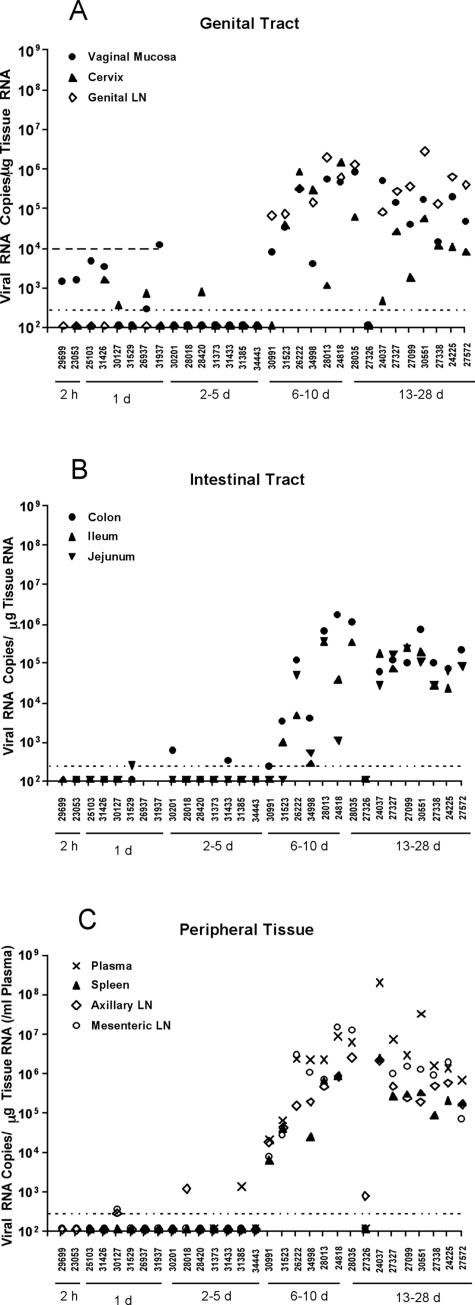
In the current global AIDS pandemic, more than half of new human immunodeficiency virus type 1 (HIV-1) infections are acquired by women through intravaginal HIV exposure. For this study, we explored pathogenesis issues relevant to the development of effective vaccines to prevent infection by this route, using an animal model in which female rhesus macaques were exposed intravaginally to a high dose of simian immunodeficiency virus (SIV). We examined in detail the events that transpire from hours to a few days after intravaginal SIV exposure through week 4 to provide a framework for understanding the propagation, dissemination, and establishment of infection in lymphatic tissues (LTs) during the acute stage of infection. We show that the mucosal barrier greatly limits the infection of cervicovaginal tissues, and thus the initial founder populations of infected cells are small. While there was evidence of rapid dissemination to distal sites, we also show that continuous seeding from an expanding source of production at the portal of entry is likely critical for the later establishment of a productive infection throughout the systemic LTs. The initially small founder populations and dependence on continuous seeding to establish a productive infection in systemic LTs define a small window of maximum vulnerability for the virus in which there is an opportunity for the host, vaccines, or other interventions to prevent or control infection.


Figures

FIG. 1.
FIG. 1.
vRNA levels in genital (A) and intestinal tract (B) tissues, peripheral lymphoid tissues (C), and plasma samples (C) of rhesus monkeys after intravaginal SIV inoculation. The bDNA assay (seeMaterials and Methods) results for the indicated tissues are displayed vertically above the animal numbers along the x axis. The animals are grouped by the timing of necropsy, as indicated by the solid horizontal lines under the animal numbers. The horizontal dotted line indicates the mean vRNA level + 2 standard deviations in the tissues of SIV-naïve monkeys and represents the cutoff value for designating positive samples (see Materials and Methods). The upper horizontal dashed line in panel A indicates the vRNA level found in the genital tract tissue of a monkey (27438) that was necropsied 24 h after intravaginal AT-2-inactivated SIVmac239 inoculation. Thus, for monkeys necropsied <48 h after intravaginal SIV inoculation, vRNA levels below this dashed line may represent virion RNA in the inoculum (see Results).
FIG. 2.
FIG. 2.
Residual virions from the inoculum trapped in cervical mucus. Sections of a cervix obtained 24 h after intravaginal exposure to SIV were cut and hybridized to SIV-specific riboprobes, and the SIV RNA signal from virions or infected cells was amplified by TSA/ELF as described in Materials and Methods. Viral RNA was not detected in cells but was detected in virions, as shown in the mucus adhering to the epithelium of the endocervical gland shown. (Left) Original magnification, ×100. The image brightness and contrast were adjusted to reveal Hoechst-counterstained nuclei and histology. (Right) Magnification, ×1,000. The image was adjusted to reveal virions at the mucosal surface.
FIG. 3.
FIG. 3.
Highly focal productive infection in an endocervix 4 days after intravaginal SIV inoculation. (A) Montage of images (magnification, ×100) of a single section of endocervix, among 20 sections examined from monkey 31385, in which one focus (encircled) of SIV RNA-positive cells was detectable. (B) SIV RNA-positive cells at a higher magnification. The double-headed arrow points to two SIV RNA-positive cells that appear to be intraepithelial lymphocytes. The single-headed arrow points to a focal collection of SIV RNA-positive cells in the endocervical mucosa. SIV RNA was detected by ISH with radiolabeled riboprobes. In the developed radioautographs viewed with transmitted light, the SIV RNA-positive cells appear black because of the large numbers of silver grains overlying the cell. Original magnification, ×100.
FIG. 4.
FIG. 4.
Relationship between duration of infection and SIV RNA levels in cells at 7 days p.i. ISH was performed with radiolabeled SIV-specific riboprobes. In developed radioautographs viewed in epipolarized light, silver grains appear white. The number of grains is proportional to the number of copies of SIV RNA per cell, which in turn is related to the length of time the cell has been infected. By this criterion, the encircled cells in the mesenteric LNs were the most recently infected. All tissues were from monkey 26222.
FIG. 5.
FIG. 5.
Frequency of SIV RNA-positive cells in mesenteric LNs. SIV RNA-positive cells detected by ISH were enumerated in sections of defined areas.
FIG. 6.
FIG. 6.
Examples of SIV RNA-positive cells in cervicovaginal and lymphatic tissues and of virions associated with follicular dendritic cells (FDCs) in LTs. SIV RNA in cells and virions was detected by ISH with 35S-labeled riboprobes. In the developed radioautographs illuminated with epipolarized light, SIV RNA-positive cells appear as bright ob-jects and FDC-associated virions appear as bright silver grains overlying follicles. (A) Cervical and vaginal tissues. On day 10, there were dense collections of SIV RNA-positive cells in inflammatory foci in the submucosa of the endocervix. The arrow points to a break in the overlying epithelium. SIV RNA-positive cells progressively decreased in frequency from days 13 to 28 and were usually associated with small foci of inflammation. (B) LNs. In mesenteric and other LNs, there were numerous SIV RNA-positive cells on day 10. Their numbers then declined. A faint signal from FDC-associated virion RNA in follicles (encircled) was detectable by day 21 and increased by day 28 in the encircled germinal center of a follicle. The red arrows each point to SIV RNA-positive cells in the adjacent T-cell zone. (C) GALT. On day 13, dense collections of SIV RNA-positive cells in the follicular aggregate of this section from the jejunum were evident in the T-cell zone (TZ; enlarged view, right panel). On day 21, FDC-associated virion RNA was detectable in the outlined germinal center (GC), in addition to SIV RNA-positive cells (right panel).
FIG. 7.
FIG. 7.
Model of delayed systemic SIV replication despite rapid dissemination. In the first 3 days p.i., the relatively few infected cells produce enough virus and infected cells to sustain the infection locally. The virus, infected cells, and DCs carrying the virus also disseminate the infection to distal sites, but the level of virus replication is insufficient to sustain a productive infection (R0 ≥ 1). On days 4 to 6 p.i., increased virus production continues at the portal of entry and now is of sufficient strength in all systemic LTs to exceed the threshold required to maintain a productive infection. Arrows indicate the direction and degree (thickness) of virus dissemination.

References

    1. Abel, K., L. Compton, T. Rourke, D. Montefiori, D. Lu, K. Rothaeusler, L. Fritts, K. Bost, and C. J. Miller. 2003. Simian-human immunodeficiency virus SHIV89.6-induced protection against intravaginal challenge with pathogenic SIVmac239 is independent of the route of immunization and is associated with a combination of cytotoxic T-lymphocyte and alpha interferon responses. J. Virol. 77:3099-3118. - PMC - PubMed
    1. Dailey, P. J., M. Zamround, R. Kelso, J. Kolberg, and M. Urdea. 1995. Quantitation of simian immunodeficiency virus (SIV) RNA in plasma of acute and chronically infected macaques using a branched DNA (bDNA) signal amplification assay. Presented at the 13th Annual Symposium on Nonhuman Primate Models of AIDS, Monterey, Calif.
    1. Fokin, A. A., F. Robicsek, and T. N. Masters. 2000. Transport of viral-size particulate matter after intravenous versus intralymphatic entry. Microcirculation 7:357-365. - PubMed
    1. Hendrickx, A. G., and M. A. Cukierski. 1987. Reproductive and developmental toxicology in nonhuman primates, p. 73-88. In C. R. Graham (ed.), Preclinical safety of biotechnology products intended for human use. Alan R. Liss, Inc., New York, N.Y. - PubMed
    1. Hirsch, V., N. Riedel, and J. I. Mullins. 1987. The genome organization of STLV-3 is similar to that of the AIDS virus except for a truncated transmembrane protein. Cell 49:307-319. - PubMed

Publication types

LinkOut - more resources