

doi: 10.1128/JVI.79.14.9315-9319.2005.
Respiratory syncytial virus nonstructural proteins NS1 and NS2 mediate inhibition of Stat2 expression and alpha/beta interferon responsiveness
Affiliations
- PMID: 15994826
- PMCID: PMC1168759
- DOI: 10.1128/JVI.79.14.9315-9319.2005
Item in Clipboard
Respiratory syncytial virus nonstructural proteins NS1 and NS2 mediate inhibition of Stat2 expression and alpha/beta interferon responsiveness
J Virol.
2005 Jul.
Abstract
Respiratory syncytial virus (RSV) subverts the antiviral interferon (IFN) response, but the mechanism for this evasion was unclear. Here we show that RSV preferentially inhibits IFN-alpha/beta signaling by expression of viral NS1 and NS2. Thus, RSV infection or expression of recombinant NS1 and NS2 in epithelial host cells causes a marked decrease in Stat2 levels and the consequent downstream IFN-alpha/beta response. Similarly, NS1/NS2-deficient RSV no longer decreases Stat2 levels or IFN responsiveness. RSV infection decreased human but not mouse Stat2 levels, so this mechanism of IFN antagonism may contribute to viral host range, as well as immune subversion.
Figures
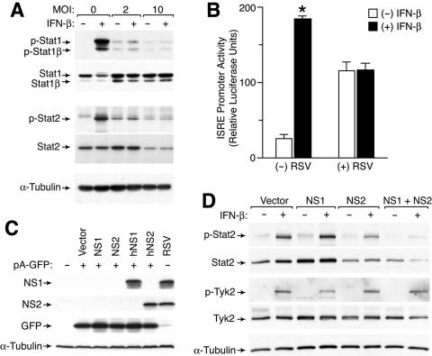
Effect of RSV infection on IFN-β responsiveness and Stat2 levels. (A) Western blot analysis for tyrosine-phosphorylated and total Stat1 and Stat2 using lysates from A549 cells that were infected with RSV Long for 24 h and then incubated with IFN-β (1,000 U/ml) for 30 min. (B) Promoter transactivation analysis from A549 cells that were transfected with an ISRE-driven luciferase reporter plasmid and a control Renilla luciferase reporter and then infected with RSV (MOI of 2) for 24 h, incubated with IFN-β (1,000 U/ml) for 12 h, and subjected to luciferase assay. Values represent luciferase light units normalized to a Renilla luciferase control (which was no different between infected and uninfected conditions), and a significant difference from the untreated condition is indicated by an asterisk. (C) Western blot analysis for NS1 and NS2 using 293T cells that were transiently transfected with 0.5 μg of pA-GFP and 1.5 μg of pcDNA5-empty, pcDNA5-NS1, pcDNA5-NS2, pcDNA5-hNS1, or pcDNA5-hNS2. Equivalent transfection efficiency was verified by flow cytometric analysis for GFP expression. RSV-infected A549 cells were included as a control. (D) Western blot analysis for phosphorylated and total Stat2 and Tyk2 using A549 cells stably expressing vector alone or hNS1, hNS2, or hNS1 plus hNS2 that were incubated with IFN-β for 30 min. Stable cell lines were generated by successive transduction first with MSCV-IRES-GFP containing no hNS, hNS1, or hNS2 insert and then with MSCV-IRES-Thy1.1 containing the appropriate hNS insert, followed by sorting for GFP and Thy1.1 expression.
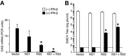
Effect of hNS1/hNS2 expression on IFN-β-dependent gene expression and viral clearance. (A) Real-time PCR analysis of 2′-5′ oligoadenylate synthetase (OAS) mRNA levels in NS-expressing A549 cells that were incubated with IFN-β (1,000 U/ml) for 6 h. Cells were transduced as described in the legend to Fig. 1. Values represent the mean ± the standard error of the mean normalized to a glyceraldehyde-3-phosphate dehydrogenase control. (B) Titers of EMCV in NS-expressing A549 cells that were treated with IFN-β (1,000 U/ml) for 12 h and then infected with EMCV (MOI of 0.1) for 24 h. Cell supernatants were used to determine EMCV titers by plaque assay. Values represent the mean ± the standard error of the mean, and dotted line indicates the lower limit of detection for the assay. Significant difference from the vector-alone condition is indicated by an asterisk.
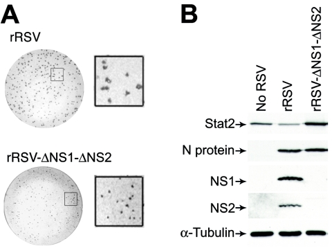
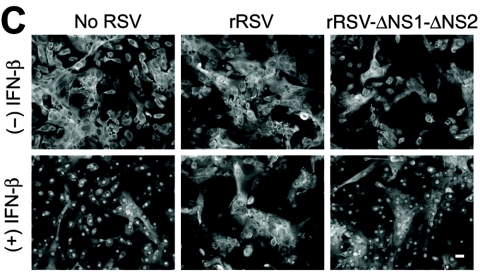
Requirement of NS1/NS2 for an RSV-dependent decrease in Stat2 expression and IFN-β responsiveness. (A) Plaque number and size for rRSV with or without NS1/NS2 (upper and lower parts, respectively) inoculated onto Vero cells for 7 days, followed by peroxidase immunostaining for RSV. Insets indicate approximately fourfold enlargement. (B) Western blot analysis for indicated proteins using hTECs that were left uninfected or infected with rRSV with or without NS1/NS2 (MOI of 10). (C) Immunofluorescence microscopy for Stat2 using hTECs that were infected with rRSV with or without NS1/NS2 (MOI of 5) for 24 h and then incubated with IFN-β (1,000 U/ml). (D) Same conditions as panel C but immunostaining for Stat2 (red, CY3 fluorescence) and RSV G protein (green, fluorescein isothiocyanate fluorescence) after incubation with IFN-β. Arrow indicates RSV-infected cell with decreased nuclear translocation of Stat2; neighboring uninfected cells and rRSV-ΔNS1-ΔNS2-infected cells exhibit normal IFN-β-driven translocation of Stat2. Bars = 20 μm.
Requirement of NS1/NS2 for an RSV-dependent decrease in Stat2 expression and IFN-β responsiveness. (A) Plaque number and size for rRSV with or without NS1/NS2 (upper and lower parts, respectively) inoculated onto Vero cells for 7 days, followed by peroxidase immunostaining for RSV. Insets indicate approximately fourfold enlargement. (B) Western blot analysis for indicated proteins using hTECs that were left uninfected or infected with rRSV with or without NS1/NS2 (MOI of 10). (C) Immunofluorescence microscopy for Stat2 using hTECs that were infected with rRSV with or without NS1/NS2 (MOI of 5) for 24 h and then incubated with IFN-β (1,000 U/ml). (D) Same conditions as panel C but immunostaining for Stat2 (red, CY3 fluorescence) and RSV G protein (green, fluorescein isothiocyanate fluorescence) after incubation with IFN-β. Arrow indicates RSV-infected cell with decreased nuclear translocation of Stat2; neighboring uninfected cells and rRSV-ΔNS1-ΔNS2-infected cells exhibit normal IFN-β-driven translocation of Stat2. Bars = 20 μm.
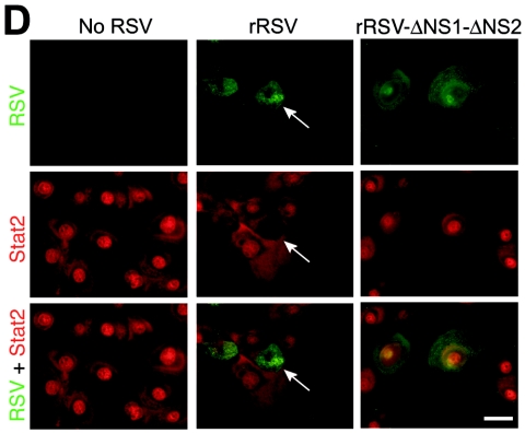
Requirement of NS1/NS2 for an RSV-dependent decrease in Stat2 expression and IFN-β responsiveness. (A) Plaque number and size for rRSV with or without NS1/NS2 (upper and lower parts, respectively) inoculated onto Vero cells for 7 days, followed by peroxidase immunostaining for RSV. Insets indicate approximately fourfold enlargement. (B) Western blot analysis for indicated proteins using hTECs that were left uninfected or infected with rRSV with or without NS1/NS2 (MOI of 10). (C) Immunofluorescence microscopy for Stat2 using hTECs that were infected with rRSV with or without NS1/NS2 (MOI of 5) for 24 h and then incubated with IFN-β (1,000 U/ml). (D) Same conditions as panel C but immunostaining for Stat2 (red, CY3 fluorescence) and RSV G protein (green, fluorescein isothiocyanate fluorescence) after incubation with IFN-β. Arrow indicates RSV-infected cell with decreased nuclear translocation of Stat2; neighboring uninfected cells and rRSV-ΔNS1-ΔNS2-infected cells exhibit normal IFN-β-driven translocation of Stat2. Bars = 20 μm.
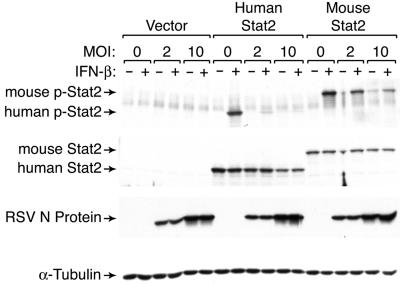
Effect of RSV infection on human versus mouse Stat2 levels. Western blot analysis for phosphorylated and total Stat2 using U6A cells stably expressing no Stat2 (vector) or human or mouse Stat2 and incubated without or with IFN-β (1,000 U/ml). Stable cell lines were generated by transduction with MSCV-IRES-GFP containing no Stat2 or human or mouse Stat2 insert, followed by sorting for GFP. Expression of control RSV N protein was not significantly different between cell lines.
References
-
- Andrejeva, J., D. F. Young, S. Goodbourn, and R. E. Randall. 2002. Degradation of STAT1 and STAT2 by the V proteins of simian virus 5 and human parainfluenza virus type 2, respectively: consequences for virus replication in the presence of alpha/beta and gamma interferons. J. Virol. 76:2159-2167. - PMC - PubMed
-
- Atreya, P. L., and S. Kulkarni. 1999. Respiratory syncytial virus strain A2 is resistant to the antiviral effects of type I interferons and human MxA. Virology 261:227-241. - PubMed
-
- Collins, P. L., M. G. Hill, E. Camargo, H. Grosfeld, R. M. Chanock, and B. R. Murphy. 1995. Production of infectious human respiratory syncytial virus from cloned cDNA confirms an essential role for the transcription elongation factor from the 5′ proximal open reading frame of the M2 mRNA in gene expression and provides a capability for vaccine development. Proc. Natl. Acad. Sci. USA 92:11563-11567. - PMC - PubMed
-
- Farrar, J. D., J. D. Smith, T. L. Murphy, S. Leung, G. R. Stark, and K. M. Murphy. 2000. Selective loss of type I interferon-induced STAT4 activation caused by a minisatellite insertion in mouse Stat2. Nat. Immunol. 1:65-69. - PubMed
Publication types
MeSH terms
Substances
LinkOut - more resources
Full Text Sources
Other Literature Sources
Miscellaneous
