Review
. 2005 Jul:(436):81-90.
doi: 10.1097/01.blo.0000171542.53342.46.

Patellofemoral joint biomechanics and tissue engineering

Affiliations
Review

Patellofemoral joint biomechanics and tissue engineering

Gerard A Ateshian et al. Clin Orthop Relat Res. 2005 Jul.

Abstract

Recent advances in the study of patellofemoral joint biomechanics have provided promising diagnosis and treatment modalities for patellofemoral joint disorders, such as quantitative assessment of cartilage lesions from noninvasive imaging, computer simulations of surgical procedures for optimizing surgical parameters and potentially predicting outcomes, and cartilage tissue engineering for the treatment of advanced degenerative joint disease. These technologies are still in development and their clinical potentials remain an ongoing topic of investigation. We review some of our progress in addressing these issues, and the important role of cartilage mechanics and lubrication in understanding the challenges regarding patellofemoral surgery and cartilage tissue engineering.


Figures

Fig 1
Fig 1
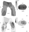
Average cartilage thickness maps for the femoral and patellar surfaces of 14 non-arthritic patellofemoral joints are shown. Reprinted with permission from Cohen ZA, Mow VC, Henry JH, Levine WN, Ateshian GA: Templates of the cartilage layers of the patellofemoral joint and their use in the assessment of osteoarthritic cartilage damage. Osteoarthritis Cartilage 11:569–579, 2003.
Fig 2
Fig 2
A–B. Average cartilage thickness maps for the femoral and patellar surfaces of 33 knees with patellofemoral joint OA are shown. (A) The mean cartilage thickness map, plotted on mean articular surface topography of OA joints is shown. (B) The difference maps against the normal template, scaled by the local standard deviation of normal cartilage thickness, and plotted on the articular surface topography from the normal template are shown. Darker regions indicate regions of cartilage loss. Reprinted with permission from Cohen ZA, Mow VC, Henry JH, Levine WN, Ateshian GA: Templates of the cartilage layers of the patellofemoral joint and their use in the assessment of osteoarthritic cartilage damage. Osteoarthritis Cartilage 11:569–579, 2003.
Fig 3
Fig 3
A–B. A physics-based model of the knee of a patient with PFJ osteoarthritis. The (A) original model and (B) the model with simulated 20 mm of tuberosity transfer are shown.
Fig 4
Fig 4
Mean and standard deviation of equilibrium tensile (square symbol) and compressive (diamond) moduli of human patellar (dashed line) and femoral (solid line) cartilage as a function of depth from the articular surface are shown. Results are from 4 men and 2 women, aged 45.5±12.0 years.
Fig 5
Fig 5
Experimental measurements of interstitial fluid load support (Wp = load supported by fluid pressure, W = total applied load) in unconfined compression stress-relaxation of cylindrical disks of human and bovine cartilage are shown. The fluid load support is higher at the articular surface, where the ratio of tensile to compressive modulus is greatest. Reprinted with permission from Park S, Krishnan R, Nicoll SB, Ateshian GA: Cartilage interstitial fluid load support in unconfined compression. J Biomech 36:1785–1796, 2003.
Fig 6
Fig 6
Cartilage friction coefficient, µeff, versus interstitial fluid load support, Wp/W, during unconfined compression creep of a cylindrical bovine cartilage disk is shown. The µeff achieves its lowest value when Wp/W is greatest. Reprinted with permission from Krishnan R, Kopacz M, Ateshian GA: Experimental verification of the role of interstitial fluid pressurization in cartilage lubrication. J Orthop Res 22:565–570, 2004.
Fig 7
Fig 7
A tissue-engineered cartilage construct in the shape of a human patellar cartilage layer, at day 35 of free-swelling culture is shown; the bottom platen is one of the two molds used for casting the chondrocyte-seeded agarose gel in the desired shape. Reprinted with permission from Hung CT, Lima EG, Mauck RL, et al: Anatomically shaped osteochondral constructs for articular cartilage repair. J Biomechanics 36:1853–1864, 2003.
Fig 8
Fig 8
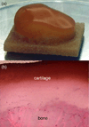
A–B. (A) Osteochondral patellar constructs, using bovine trabecular bone as the substrate, are shown. (B) A section of osteochondral construct after 35 days of free-swelling culture shows GAG distribution (stain, Safranin-O; magnification 4×).

References

    1. Adam C, Eckstein F, Milz S, et al. The distribution of cartilage thickness in the knee-joints of old-aged individuals -- measurement by A-mode ultrasound. Clin Biomech (Bristol, Avon) 1998;13:1–10. - PubMed
    1. Ahmed AM, Burke DL. In-vitro measurement of static pressure distribution in synovial joints--Part I: Tibial surface of the knee. J Biomech Eng. 1983;105:216–225. - PubMed
    1. Ahmed AM, Burke DL, Yu A. In-vitro measurement of static pressure distribution in synovial joints--Part II: Retropatellar surface. J Biomech Eng. 1983;105:226–236. - PubMed
    1. Akizuki S, Mow VC, Muller F, et al. Tensile properties of human knee joint cartilage: I. Influence of ionic conditions, weight bearing, and fibrillation on the tensile modulus. J Orthop Res. 1986;4:379–392. - PubMed
    1. Armstrong CG, Mow VC. Variations in the intrinsic mechanical properties of human articular cartilage with age, degeneration, and water content. J Bone Joint Surg Am. 1982;64:88–94. - PubMed

Publication types

MeSH terms