Review
. 2005 Jul;6(7):533-43.
doi: 10.1038/nrg1637.

Synthetic biology

Affiliations
Review

Synthetic biology

Steven A Benner et al. Nat Rev Genet. 2005 Jul.

Abstract

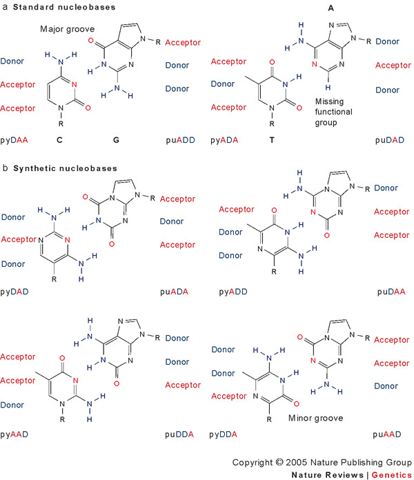
Synthetic biologists come in two broad classes. One uses unnatural molecules to reproduce emergent behaviours from natural biology, with the goal of creating artificial life. The other seeks interchangeable parts from natural biology to assemble into systems that function unnaturally. Either way, a synthetic goal forces scientists to cross uncharted ground to encounter and solve problems that are not easily encountered through analysis. This drives the emergence of new paradigms in ways that analysis cannot easily do. Synthetic biology has generated diagnostic tools that improve the care of patients with infectious diseases, as well as devices that oscillate, creep and play tic-tac-toe.


Conflict of interest statement

The authors declare no competing financial interests.

Figures

Figure 1
Figure 1. Examples of alternative nucleobases.
Parts of the nucleobases of DNA can be used as interchangeable building modules. The blue units are the hydrogen bonding donor (D) collections of atoms. The red units are the hydrogen bonding acceptor (A) collections of atoms. a | The four standard nucleobases are shown. b | Shuffling the hydrogen bond donor and acceptor modules generates eight additional nucleotides, which constitute a synthetic genetic system. These synthetic bases have been used in an artificial genetic system that can support Darwinian evolution. A, adenine; C, cytosine; G, guanine; Pu, purine; Py, pyrimidine; T, thymine.
Figure 2
Figure 2. Branched DNA assay developed by scientists at Chiron and Bayer Diagnostics.
The target RNA molecule to be detected (the analyte) is attached to the plastic of a microwell (bottom) by the hybridization of the analyte to a series of capture probes. This complex then captures, through hybridization, a target probe, which in turn hybridizes to a pre-amplifier molecule, thereby 'sandwiching' the analyte between the capture probe and the pre-amplifier. The pre-amplifier captures a branched DNA dendrimer (amplifier) that contains several signalling molecules on each branch. As a consequence of the branching, a single analyte assembles a large number of signalling molecules in the microwell. These assays use the expanded genetic alphabet shown in Fig. 1. When standard nucleotides were used to assemble the signalling nanostructure, significant noise was seen, because non-target DNA that was present in the biological sample was captured by the probes in the microwell even in the absence of analyte. Incorporating components of the artificial genetic alphabet in the dendrimer reduced the noise. As a consequence, the assay now helps manage the care of some 400,000 patients annually, detecting as few as eight molecules of the analyte DNA in a sample.
Figure 3
Figure 3. A self-templating system built from peptide units.
a | A de novo designed peptide ligase. α-helical peptides A and B bind to the electrostatically complementary α-helical peptide C to form the C·A·B ternary complex, which is composed of two coiled coils. Peptide A has a modified amino terminus that reacts with the chemically modified carboxyl terminus of peptide B on formation of the ternary complex. The reaction of peptides A and B is a ligation that forms the product C·P (C·P* represents the chemical reaction between A and B to produce P). b | Peptide replicator schematic based on the reaction illustrated in part a, showing the reaction of peptide A with peptide B on formation of a ternary complex with peptide C. Peptides AL, BL, and CLL are composed of L-amino acids, whereas peptides AD, BD, and CDD are composed of D-amino acids. Peptides CLL and CDD are produced autocatalytically in a template-directed fashion through the reaction of precursors AL with BL, and AD with BD, respectively. Therefore, this replicator is stereochemically selective, only producing products (CLL and CDD) that are isomerically pure. Part b modified, with permission, from Ref. © American Chemical Society (2001) and from Nature Ref. © (2001) Macmillan Magazines Ltd.
Figure 4
Figure 4. Using proteins as interchangeable parts in synthetic biology.
a | The combination of enzymes from three sources in a Ralstonia eutropha host generated a strain that produced large amounts of a poly(3-hydroxybutyrate-co-3-hydroxyhexanoate) polymer from fructose. All enzymes from Ralstonia eutropha are shown in black, whereas those from Areomonas caviae and Streptomyces cinnamonensis are shown in green and red, respectively. Modified, with permission, from Ref. © American Chemical Society (2002). b | The combination of enzymes from three sources in an Escherichia coli host generated a strain of the bacterium that produced a precursor for artemisinin, an antimalarial drug. The challenge of this experiment lay in the need to curtail the pathway to recognize and avoid metabolite toxicity, while optimizing the yield of the desired product. The general methodology for the pathway design was to use an engineered mevalonate pathway that is absent in E. coli, rather than the DXP (1-deoxy-D-xylulose 5-phosphate) pathway that is native to the organism. The synthetic operons used are depicted, and the engineered pathway metabolites are shown in red. In the engineered mevalonate pathway, the fan of genes from ERG12 to ispA exist on multiple plasmids to tune the pathway for optimization of the product while avoiding metabolite toxicity. As depicted at the bottom of the figure, the E. coli strain DYM1, a strain deficient in isoprenoid synthesis, was used, because the DXP pathway was found to limit product yield, probably owing to an unrecognized link between the pathway and physiological control elements in the organism. Enzymes used (isolated from Saccharomyces cerevisiae unless otherwise noted): ADS, amorphadiene synthase; atoB, acetoacetyl-CoA thiolase (E. coli); dxs, 1-deoxy-D-xylulose 5-phosphate synthase; ERG12, mevalonate kinase; ERG8, phosphomevalonate kinase; HMGS, HMG-CoA synthase; idi, IPP isomerase (E. coli); ippHp, IPP isomerase (H. pluvialis); ispA, FPP synthase (E. coli); ispC, 1-deoxy-D-xylulose 5-phosphate reductoisomerase; MVD1, mevalonate pyrophosphate decarboxylase; tHMGR, truncated HMG-CoA reductase. Pathway intermediates: AA-CoA, acetoacetly-CoA; A-CoA, acetyl-CoA; CDP-Me, 4-diphosphocytidyl-2-C-methyl-D-erythritol; CDP-ME2P, 4-diphosphocytidyl-2-C-methyl-D-erythritol 2-phosphate; DMAPP, dimethylallyl pyrophosphate; DXP, 1-deoxy-D-xylulose 5-phosphate; FPP, farnesyl pyrophosphate; G3P, glyceraldehyde 3-phosphate; HMB4PP, 1-hydroxy-2-methyl-2-(E)-butenyl 4-pyrophosphate; HMG-CoA, hydroxymethylglutaryl-CoA; IPP, isopentenyl pyrophosphate; Mav-P, mevalonate 5-phosphate; ME-2,4cPP, 2-C-methyl-D-erythritol 2,4-cyclopyrophosphate; MEP, 2-C-methyl-D-erythritol 4-phosphate; Mev-PP, mevalonate pyrophosphate. Adapted, with permission, from Nature Biotechnology Ref. © (2003) Macmillan Magazines Ltd.
Figure 5
Figure 5. The design and application of the repressilator.
a | Schematic showing the regulation pattern that forms the basis of a repressilator. Three gene–promoter pairs are arranged so that the product derived from the expression of the gene following a promoter is a repressor for the next promoter in the cycle. Black connecting lines show that promoter PLlacO1 controls the transcription of the gene tetR-lite, the tetracycline repressor protein TetR represses PLtetO1, which is the next promoter in the sequence. PLtetO1 in turn controls the transcription of cI-lite, and the protein CI represses the promoter PR. Finally, PR controls the expression of lacI-lite, and the lactose repressor protein LacI represses PLlacO1, completing the cycle. The suffix '-lite' refers to the presence of tags that increase the degradation rate of the proteins. b | The luminescence pattern of a reporter plasmid that carries GFP under the transcriptional control of the PLtetO1 promoter, when the reporter construct is transferred to an Escherichia coli in the presence of the repressilator. As the experimental trace shows, the oscillation of the TetR repressor expressed from the repressilator results in the time dependent oscillation of GFP expression. Bars at the bottom of the diagram show the timing of cell division events. The period of the oscillations is longer than the cell division time, and the cycle of oscillations continues in the subsequent generations. Adapted, with permission, from Nature Ref. © (2000) Macmillan Magazines Ltd.

References

    1. Hobom B. Surgery of genes. At the doorstep of synthetic biology. Medizin. Klinik. 1980;75:14–21. - PubMed
    1. Rawls, R. 'Synthetic Biology' makes its debut. Chem. Eng. News 49–53 (24 April 2000).
    1. Benner SA. Redesigning life. Organic chemistry and the evolution of protein. Chimia. 1987;41:142–148.
    1. Szostak JW, Bartel DP, Luisi PL. Synthesizing life. Nature. 2001;409:387–390. doi: 10.1038/35053176. - DOI - PubMed
    1. Benner, S. A. Act natural. Nature421, 118 (2003). This paper outlines the importance of pursuing the science of synthetic biology. - PubMed