Review
. 2005 Jul;6(7):533-44.
doi: 10.1038/nrn1704.

Pain and emotion interactions in subregions of the cingulate gyrus

Affiliations
Review

Pain and emotion interactions in subregions of the cingulate gyrus

Brent A Vogt. Nat Rev Neurosci. 2005 Jul.

Abstract

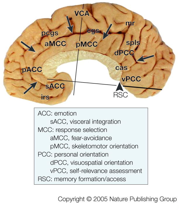
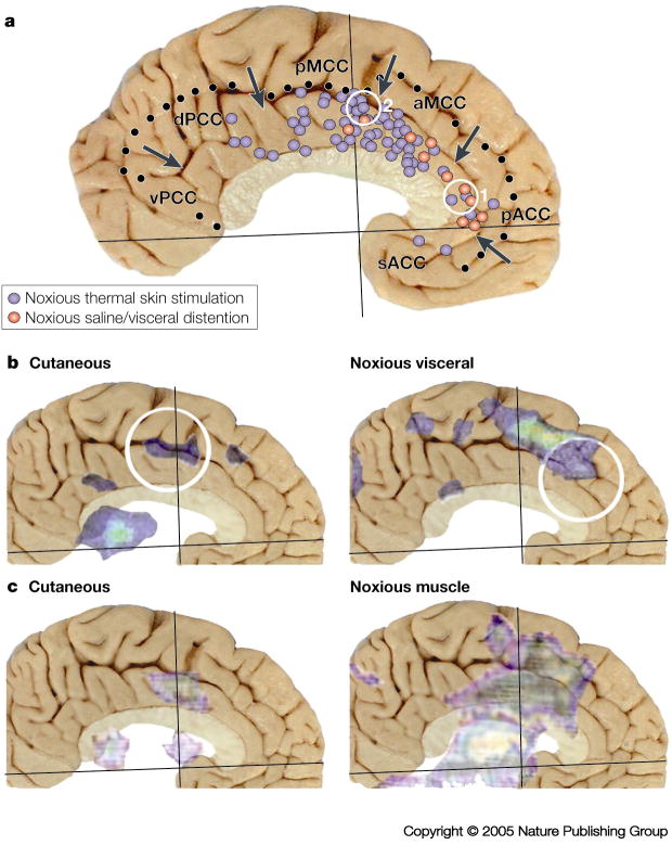
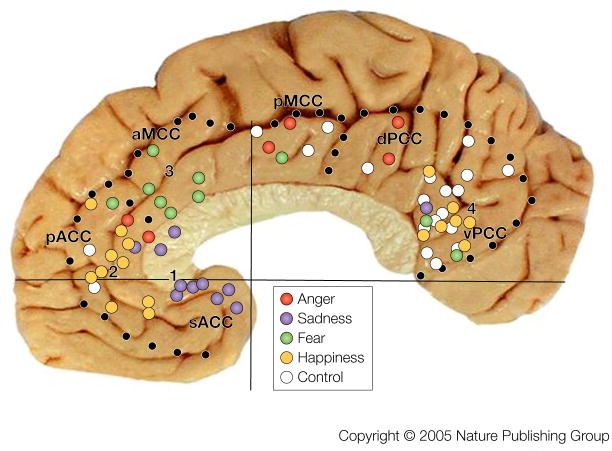
Acute pain and emotion are processed in two forebrain networks, and the cingulate cortex is involved in both. Although Brodmann's cingulate gyrus had two divisions and was not based on any functional criteria, functional imaging studies still use this model. However, recent cytoarchitectural studies of the cingulate gyrus support a four-region model, with subregions, that is based on connections and qualitatively unique functions. Although the activity evoked by pain and emotion has been widely reported, some view them as emergent products of the brain rather than of small aggregates of neurons. Here, we assess pain and emotion in each cingulate subregion, and assess whether pain is co-localized with negative affect. Amazingly, these activation patterns do not simply overlap.


Figures

Box 1
Box 1
Box 2
Box 2
Figure 1
Figure 1. Distribution of the Four Cingulate Regions & Subregions
Borders are marked with arrows and were determined in this and six other postmortem cases that were coregistered to a stereotaxic atlas with the vertical plane at the anterior commissure (VCA) and the anterior-posterior commissural line. A functional overview derived from many literature analyses is provided with general regional function along with subregional specializations where known. Abbreviations: cas, callosal sulcus; cgs, cingulate sulcus; irs, inferior rostral sulcus; mr, marginal ramus of cgs; pcgs, paracingulate sulcus.
Figure 2
Figure 2. Nociceptive Cingulate Neurons
Medial surface of the rabbit brain showing its cingulate regions (ACC, MCC, RSC) and the location of a high density of nociceptive neurons (black) and a low-moderate density of such neurons (grey). (The rabbit does not have PCC areas 23 and 31 like primates.) The rasters display neuron spike discharges for two neurons and the arrows show stimulus onset (↑) and offset (↓). Noxious mechanical stimulation with a serrated forceps evoked a response of the neuron on the left regardless of where the stimulus was applied to the skin and demonstrates that no one neuron in ACC can determine where on the body a noxious stimulus is located. The neuron on the right is a multimodal nociceptive neuron because it responds briskly to both noxious mechanical and heat stimulation. Although the unit did respond to tap stimulation, light brushing of the skin (open arrows) failed to evoke a response. These multimodal nociceptive units provide little information about the characteristics of particular noxious stimuli.
Figure 3
Figure 3. Human imaging during acute nociceptive stimulation
A. Summary of peak activation sites in 40 studies during noxious thermal stimulation of the skin and noxious hypertonic saline or visceral distention (References provided online). Cutaneous activations were almost homogeneous throughout MCC with somewhat fewer in the rostral part of aMCC and most in the dorsal and rostral part of pMCC, while visceral activity was greatest in pACC and some in rostral aMCC. The two yellow circles are for activations associated with the opioid placebo (1.55) and the acupuncture placebo (2.57). B./C. Activation sites for two studies were coregistered to the postmortem control case because two different noxious stimulation paradigms were applied to the same subjects providing a more accurate differentiation of the topography of activation sites. As a rule, noxious cutaneous stimulation evoked small and more caudal activity in pMCC, while noxious esophageal distention (B.36) or electrical stimulation of muscle (C.37) evoked larger and more rostral activity in aMCC. Differential activation of parts of MCC likely reflects the differential recruitment of the caudal and rostral cingulate motor areas and their associated cognitive functions.
Figure 4
Figure 4. Nociceptive afferents to cingulate cortex
Three sources of nociceptive inputs to “Pf” arise from lamina I of the spinal cord, subnucleus reticularis dorsalis (SRD) and the parabrachial nucleus (PB). The four asterisks along the medial surface of the monkey brain show the levels at which the thalamic, PB, SRD, and high cervical spinal cord sections were photographed. These are immunohistochemical sections for an antibody to microtubule associated protein 2 (MAP2) which label neuron dendrites and they were counterstained with thionin because some neurons do not express this antigen. The Pf nucleus is enclosed in quotation marks because it is a representative of the MITN of which there are many nuclei that receive nociceptive spinothalamic inputs and project in turn to cingulate cortex (see BOX #2). We believe the greatest density of nociceptive inputs is to aMCC as shown with the large arrow, while more modest projections are to ACC and pMCC. Abbreviations: CM, centre medianum thalamic nucleus; EC, external cuneate nucleus; Gi, gigantocellular reticular nucleus; IO, inferior olive; LC, locus coeruleus; LD, laterodorsal thalamic nucleus; MD, mediodorsal thalamic nucleus; scp, superior cerebellar peduncle; SpV, spinal nucleus and tract of the trigeminal nerve; VPL, ventral posterolateral thalamic nucleus.
Figure 5
Figure 5. Cingulate Emotion Processing
Peak activation sites from 23 studies during simple emotions in the context of the regionalized cingulate gyrus (references provided online). Four groups of active sites are numbered and control conditions with non-emotional scripts and faces are coded with white dots. Each numbered aggregate of sites is located in a different subregion and this suggests a different role in processing of emotional information; i.e., a different relevance to autonomic integration, skeletomotor output, and personal orientation as predicted by the four-region, neurobiological model.

References

    1. Aggleton JP. The Amygdala. Oxford University Press; New York: 2001.
    1. Davidson RJ, Scherer KR, Goldsmith HH. Handbook of Affective Sciences. Oxford University Press; New York: 2003.
    1. Melzack R, Casey KL. Sensory, motivational and central control determinants of pain. A new conceptual model. In: Kenshalo DR, editor. The Skin Senses. Springfield, IL: Thomas; 1968. pp. 423–439.
    1. Derbyshire SWG. Exploring the pain neuromatrix. Curr Rev Pain. 2000;6:467–477. - PubMed
    1. Peyron R, Laurent B, Garcia-Larrea L. Functional imaging of brain responses to pain: A review and meta-analysis. Neurophysiol Clin. 2000;30:263–288. - PubMed

Publication types