. 2005 Aug;141(2):238-47.
doi: 10.1111/j.1365-2249.2005.02849.x.

Bacterial lipopolysaccharide both renders resistant mice susceptible to mercury-induced autoimmunity and exacerbates such autoimmunity in susceptible mice

Affiliations

Bacterial lipopolysaccharide both renders resistant mice susceptible to mercury-induced autoimmunity and exacerbates such autoimmunity in susceptible mice

M Abedi-Valugerdi et al. Clin Exp Immunol. 2005 Aug.

Abstract

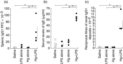
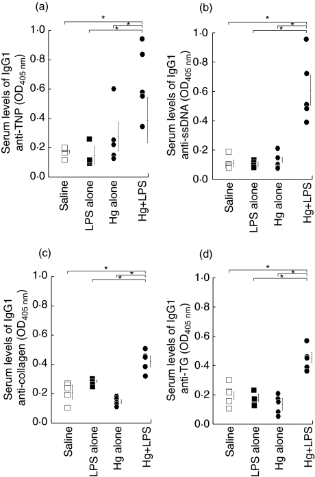
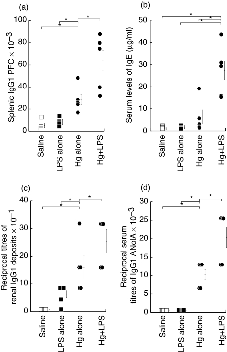
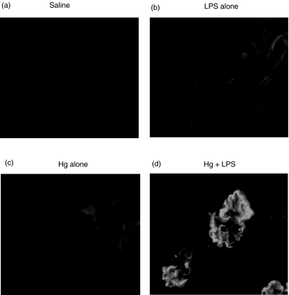
The initiation and severity of systemic autoimmune diseases are influenced by a variety of genetic and environmental factors, in particular bacterial infections and products. Here, we have employed bacterial lipopolysaccharide (LPS), which non-specifically activates the immune system, to explore the involvement of innate immunity in mercury-induced autoimmunity in mice. Following treatment of mouse strains resistant [DBA/2 (H-2(d))] or susceptible [SJL(H-2(s))] to such autoimmunity with mercuric chloride and/or LPS or with physiological saline alone (control), their immune/autoimmune responses were monitored. Resistant DBA/2 mice were rendered susceptible to mercury-induced autoimmunity by co-administration of LPS, exhibiting pronounced increases in the synthesis of IgG1 and IgE, high titres of IgG1 deposits in the kidneys and elevated circulating levels of IgG1 antibodies of different specificities. Furthermore, the percentages of the T cells isolated from the spleens of DBA/2 mice exposed to both mercury and LPS that produced pro-inflammatory cytokines were markedly increased by in vitro stimulation with phorbol myristate acetate (PMA) and ionomycin, which was not the case for splenic T cells isolated from mice receiving mercuric chloride, LPS or saline alone. In addition, exposure of susceptible SJL mice to mercury in combination with LPS aggravated the characteristic features of mercury-induced autoimmunity, including increased synthesis of IgG1 and IgE, the production of IgG1 anti-nucleolar antibodies (ANolA) and the formation of renal deposits of IgG1. In summary, our findings indicate that activation of the innate immune system plays a key role in both the induction and severity of chemically induced autoimmunity.


Figures

Fig. 1
Fig. 1
Administration of bacterial lipopolysaccharide (LPS) during treatment with mercuric chloride renders DBA/2 mice susceptible to mercury-induced autoimmunity. Group 1 (five mice per group) (filled hexagons) received repeated subcutaneous injections of subtoxic doses of mercuric chloride (Hg) for a period of 4 weeks. In addition to similar administration of mercuric chloride, group 2 (filled circles) was injected i.v. with LPS (50 µg per mouse) on the 1st and 15th days of mercury treatment. The two control groups 3 (filled squares) and 4 (open squares) received LPS alone on two occasions or repeated injections of physiological saline, according to the corresponding schedules used for groups 2 and 1, respectively. At the end of the period of treatment (a) the percentages of splenic cells secreting IgG1 antibodies (PFC) were determined employing the protein A plaque assay. In addition (b) the levels of IgE in the serum samples were determined employing a sandwich enzyme-linked immunosorbent assay (ELISA) assay and (c) the kidneys were analysed for the presence of glomerular deposits of IgG1 utilizing a direct immunofluorescence technique, as described in the Materials and methods. The mean ± s.e. values are presented as thin vertical lines. *P < 0·05 for the difference between the two groups indicated. These results are representative of three independent experiments.
Fig. 3
Fig. 3
Administration of bacterial lipopolysaccharide (LPS) together with mercuric chloride to DBA/2 mice increases their production of IgG1 antibodies to both exogenous and endogenous antigens. The levels of IgG1 antibodies directed against TNP (a), ssDNA (b), collagen (c) and thyroglobulin (d) in serum samples from the same animals presented in Fig. 1 were determined employing sandwich enzyme-linked immunosorbent assay (ELISA) assays, as described in detail in the Materials and methods. In all cases a serum dilution of 1 : 40 was utilized and the values obtained were within the linear range of the dilution curve. The mean ± s.e. values are presented as thin vertical lines. *P < 0·05 for the difference between the two groups indicated. These results are representative of two independent experiments.
Fig. 4
Fig. 4
Exposure to bacterial lipopolysaccharide (LPS) potentiates the development of mercury-induced autoimmunity in susceptible SJL mice. This experiment was the same as that described in the legend to Fig. 1, except that SJL mice were used and serum levels of anti-nucleolar autoantibodies (ANolA) (d) were also determined, employing an indirect immunofluorescence procedure. The mean ± s.e. values are presented as thin vertical lines. *P < 0·05 for the difference between the two groups indicated. These results are representative of three independent experiments.
Fig. 2
Fig. 2
Immunofluorescent detection of glomerular deposits of IgG1 in the kidneys of DBA/2 mice administered mercuric chloride in combination with lipopolysaccharide (LPS). The kidneys from the same animals presented in Fig. 1 were tested for the presence of glomular deposits of IgG1 employing the direct immunofluorescence procedure described in the Materials and methods. Representative kidney sections from animals administered saline (a), LPS (b) or mercury (c) alone show no green fluorescence intensity; whereas the kidney of a DBA/2 mouse treated with mercury in combination with LPS (d) displays intense green fluorescence in the glomeruli.

Similar articles

Cited by

References

    1. Brickman CM, Shoenfeld Y. The mosaic of autoimmunity. Scand J Clin Lab Invest Suppl. 2001;235:3–15. - PubMed
    1. Luppi P, Rossiello MR, Faas S, Trucco M. Genetic background and environment contribute synergistically to the onset of autoimmune diseases. J Mol Med. 1995;73:381–93. - PubMed
    1. Mok CC, Lau CS. Pathogenesis of systemic lupus erythematosus. J Clin Pathol. 2003;56:481–90. - PMC - PubMed
    1. Zandman-Goddard G, Shoenfeld Y. SLE and infections. Clin Rev Allergy Immunol. 2003;25:29–40. - PubMed
    1. Adelman MK, Marchalonis JJ. Endogenous retroviruses in systemic lupus erythematosus: candidate lupus viruses. Clin Immunol. 2002;102:107–16. - PubMed

Publication types