. 2005 Aug;141(2):248-60.
doi: 10.1111/j.1365-2249.2005.02835.x.

Hypoxia aggravates lipopolysaccharide-induced lung injury

Affiliations

Hypoxia aggravates lipopolysaccharide-induced lung injury

D Vuichard et al. Clin Exp Immunol. 2005 Aug.

Abstract

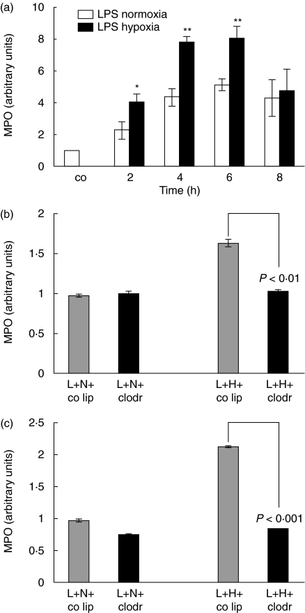
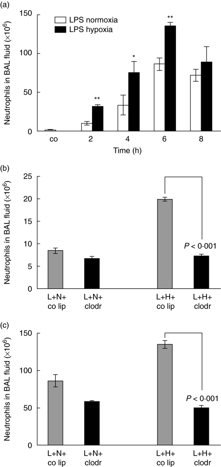
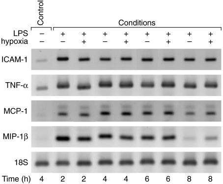
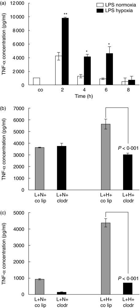
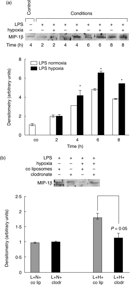
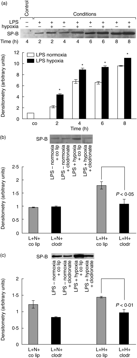
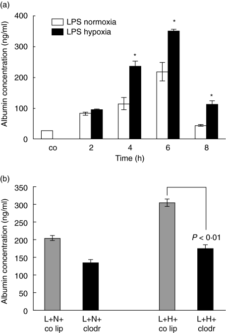
The animal model of inflammatory response induced by intratracheal application of lipopolysaccharide includes many typical features of acute lung injury or the acute respiratory distress syndrome. A number of experimental investigations have been performed to characterize the nature of this injury more effectively. In inflammatory conditions, hypoxia occurs frequently before and in parallel with pulmonary and non-pulmonary pathological events. This current study was designed to examine the in vivo effect of hypoxia as a potentially aggravating condition in endotoxin-induced lung injury. Lipopolysaccharide, 150 microg, was instilled intratracheally into rat lungs, and thereafter animals were exposed to either normoxia or hypoxia (10% oxygen). Lungs were collected 2, 4, 6 and 8 h later. Inflammatory response and tissue damage were evaluated by quantitative analysis of inflammatory cells and mediators, surfactant protein and vascular permeability. A significantly enhanced neutrophil recruitment was seen in lipopolysaccharide-animals exposed to hypoxia compared to lipopolysaccharide-animals under normoxia. This increased neutrophil accumulation was triggered by inflammatory mediators such as tumour necrosis factor-alpha and macrophage inflammatory protein-1beta, secreted by alveolar macrophages. Determination of vascular permeability and surfactant protein-B showed enhanced concentrations in lipopolysaccharide-lungs exposed to hypoxia, which was absent in animals previously alveolar macrophage-depleted. This study demonstrates that hypoxia aggravates lipopolysaccharide injury and therefore represents a second hit injury. The additional hypoxia-induced inflammatory reaction seems to be predominantly localized in the respiratory compartment, underlining the compartmentalized nature of the inflammatory response.


Figures

Fig. 1
Fig. 1
(a) Determination of myeloperoxidase (MPO) activity as a measure of parenchymal neutrophil content. Lipopolysaccharide (LPS), 150 µg, was instilled intratracheally and animals were exposed to normoxia or hypoxia for 2, 4, 6 and 8 h. Lungs were lavaged; the right lung of each animal was removed and homogenized in sample buffer. Optical density was measured at 420 nm over 360 s. The value for control animals was defined as 1 and all other values were adapted. Values are mean ± s.e.m. from five animals. *P < 0·05, **P < 0·01 between LPS-normoxia and LPS-hypoxia animals. (b) Determination of MPO activity as a measure of parenchymal neutrophil content. Animals were pretreated with control liposomes (co lip) or clodronate-liposomes (clodr); 72 h later 150 µg lipopolysaccharide (L) was instilled intratracheally and animals were exposed to either normoxia (N) or hypoxia (H) for 2 h. Values are mean ± s.e.m. from five animals. (c) Determination of myeloperoxidase (MPO) activity as a measure of parenchymal neutrophil content. Animals were pretreated with control liposomes (co lip) or clodronate-liposomes (clodr); 72 h later 150 µg lipopolysaccharide (L) was instilled intratracheally and animals were exposed to either normoxia (N) or hypoxia (H) for 6 h. Values are mean ± s.e.m. from five animals.
Fig. 2
Fig. 2
(a) Total cell count in bronchoalveolar lavage (BAL) fluid. Lipopolysaccharide (LPS), 150 µg, was instilled intratracheally and animals were exposed to normoxia or hypoxia for 2, 4, 6 and 8 h. Lungs were lavaged and cells from BAL fluid were analysed using cytospins and Diff-Quick staining. Values are mean ± s.e.m. from five animals. *P < 0·05 and **P < 0·01 between LPS-normoxia and LPS-hypoxia animals. (b) Total cell count in BAL fluid. Animals were pretreated with control liposomes (co lip) or clodronate-liposomes (clodr); 72 h later 150 µg LPS (L) was instilled intratracheally and animals were exposed to either normoxia (N) or hypoxia (H) for 2 h. Values are mean ± s.e.m. from five animals. (c) Total cell count in BAL fluid. Animals were pretreated with control liposomes (co lip) or clodronate-liposomes (clodr); 72 h later 150 µg lipopolysaccharide (L) was instilled intratracheally and animals were exposed to either normoxia (N) or hypoxia (H) for 6 h. Values are mean ± s.e.m. from five animals.
Fig. 3
Fig. 3
Changes in expression of intercellular adhesion molecule-1 (ICAM-1), tumour necrosis factor-α (TNF-α), monocyte chemoattractant protein-1 (MCP-1) and macrophage inflammatory protein-1β (MIP-1β) mRNA in lipopolysaccharide (LPS)-normoxia and LPS-hypoxia animals. LPS, 150 µg, was instilled intratracheally and animals were exposed to normoxia or hypoxia for 2, 4, 6 and 8 h. Lungs were lavaged, and whole lung RNA was extracted. Reverse transcription-polymerase chain reaction (RT-PCR) was performed according to the primers and annealing thermocycle conditions shown in Table 1. Equal loading was shown with 18S bands.
Fig. 4
Fig. 4
(a) Tumour necrosis factor-α (TNF-α) protein concentration in bronchoalveolar lavage (BAL) fluid of lipopolysaccharide (LPS)-normoxia and LPS-hypoxia animals. LPS, 150 µg, was instilled intratracheally and animals were exposed to normoxia or hypoxia for 2, 4, 6 and 8 h. Lungs were lavaged, and TNF-α was determined using a standard enzyme-linked immunosorbent assay (ELISA). Values are mean ± s.e.m. from five animals. *P < 0·05 and **P < 0·01 between LPS-normoxia and LPS-hypoxia animals. (b) TNF-α protein concentration in BAL fluid of alveolar macrophage-competent and -depleted animals, exposed to lipopolysaccharide (LPS)-normoxia and LPS-hypoxia. Animals were pretreated with control liposomes (co lip) or clodronate-liposomes (clodr); 72 h later 150 µg LPS (L) was instilled intratracheally and animals were exposed to either normoxia (N) or hypoxia (H) for 2 h. TNF-α was determined using a standard ELISA. Values are mean ± s.e.m. from five animals. (c) TNF-α protein concentration in BAL fluid of alveolar macrophage-competent and -depleted animals, exposed to LPS-normoxia and LPS-hypoxia. Animals were pretreated with control liposomes (co lip) or clodronate-liposomes (clodr); 72 h later 150 µg LPS (L) was instilled intratracheally and animals were exposed to either normoxia (N) or hypoxia (H) for 6 h. TNF-α was determined using a standard ELISA. Values are mean ± s.e.m. from five animals.
Fig. 5
Fig. 5
(a)Macrophage inflammatory protein-1β (MIP-1β) protein determination in bronchoalveolar lavage (BAL) fluid of lipopolysaccharide (LPS)-normoxia and LPS-hypoxia animals. LPS, 150 µg, was instilled intratracheally and animals were exposed to normoxia or hypoxia for 2, 4, 6 and 8 h. Lungs were lavaged, and proteins were electrophoresed on a sodium dodecyl sulphate-polyacrylamide gel and transblotted to a nitrocellulose membrane. The blot represents one of five experiments. Densitometry was performed. Value for control animals was defined as 1 and all other values were adapted. Values are mean ± s.e.m. from five animals. *P < 0·05 between LPS-normoxia and LPS-hypoxia animals. (b) MIP-1β protein concentration in BAL fluid of alveolar macrophage-competent and -depleted animals, exposed to LPS-normoxia and LPS-hypoxia. Animals were pretreated with control liposomes (co lip) or clodronate-liposomes (clodr); 72 h later 150 µg LPS (L) was instilled intratracheally and animals were exposed to either normoxia (N) or hypoxia (H) for 6 h. BAL fluid proteins were electrophoresed on a SDS-polyacrylamide gel and transblotted to a nitrocellulose membrane. The blot represents one of five experiments.
Fig. 6
Fig. 6
(a) Surfactant protein-B (SP-B) protein determination in bronchoalveolar lavage (BAL) fluid of lipopolysaccharide (LPS)-normoxia and LPS-hypoxia animals. LPS, 150 µg, was instilled intratracheally and animals were exposed to normoxia or hypoxia for 2, 4, 6 and 8 h. Lungs were lavaged, and proteins were electrophoresed on a sodium dodecyl sulphate-polyacrylamide gel and transblotted to a nitrocellulose membrane. The blot represents one of five experiments. Densitometry was performed. Value for control animals was defined as 1 and all other values were adapted. Values are mean ± s.e.m. from five animals. *P < 0·05 between LPS-normoxia and LPS-hypoxia animals. (b) SP-B protein concentration in BAL fluid of alveolar macrophage-competent and -depleted animals, exposed to LPS-normoxia and LPS-hypoxia. Animals were pretreated with control liposomes (co lip) or clodronate-liposomes (clodr); 72 h later 150 µg LPS (L) was instilled intratracheally and animals were exposed to either normoxia (N) or hypoxia (H) for 2 h. BAL fluid proteins were electrophoresed on a sodium dodecyl sulphate-polyacrylamide gel and transblotted to a nitrocellulose membrane. The blot represents one of five experiments. (c) SP-B protein concentration in BAL fluid of alveolar macrophage-competent and -depleted animals, exposed to LPS-normoxia and LPS-hypoxia. Animals were pretreated with control liposomes (co lip) or clodronate-liposomes (clodr); 72 h later 150 µg LPS (L) was instilled intratracheally and animals were exposed to either normoxia (N) or hypoxia (H) for 6 h. BAL fluid proteins were electrophoresed on a sodium dodecyl sulphate-polyacrylamide gel and transblotted to a nitrocellulose membrane. The blot represents one of five experiments.
Fig. 7
Fig. 7
Enzyme-linked immunosorbent assay (ELISA) for albumin content in bronchoalveolar lavage (BAL) fluid. Lipopolysaccharide (LPS), 150 µg, was instilled intratracheally and animals were exposed to normoxia or hypoxia for 2, 4, 6 and 8 h. Lungs were lavaged and albumin was analysed in BAL fluid. Values are means ± s.e.m. from five animals. *P < 0·05 between LPS-normoxia and LPS-hypoxia animals. (b) ELISA for albumin content in BAL fluid of alveolar macrophage-competent and -depleted animals, exposed to LPS-normoxia and LPS-hypoxia. Animals were pretreated with control liposomes (co lip) or clodronate-liposomes (clodr); 72 h later 150 µg LPS (L) was instilled intratracheally and animals were exposed to either normoxia (N) or hypoxia (H) for 6 h. The blot represents one of five experiments.

Comment in

Similar articles

Cited by

References

    1. Ulich TR, Yin S, Remick DG, Russell D, Eisenberg SP, Kohno T. Intratracheal administration of endotoxin and cytokines. IV. The soluble tumor necrosis factor receptor type I inhibits acute inflammation. Am J Pathol. 1993;142:1335–8. - PMC - PubMed
    1. Ulich TR, Howard SC, Remick DG, et al. Intratracheal administration of endotoxin and cytokines. VIII. LPS induces E-selectin expression; anti-E-selectin and soluble E-selectin inhibit acute inflammation. Inflammation. 1994;18:389–98. - PubMed
    1. Ulich TR, Howard SC, Remick DG, et al. Intratracheal administration of endotoxin and cytokines. VI. Antiserum to CINC inhibits acute inflammation. Am J Physiol. 1995;268:L245–50. - PubMed
    1. Beck-Schimmer B, Schimmer RC, Warner RL, et al. Expression of lung vascular and airway ICAM-1 after exposure to bacterial lipopolysaccharide. Am J Respir Cell Mol Biol. 1997;17:344–52. - PubMed
    1. van Helden HP, Kuijpers WC, Steenvoorden D, et al. Intratracheal aerosolization of endotoxin (LPS) in the rat: a comprehensive animal model to study adult (acute) respiratory distress syndrome. Exp Lung Res. 1997;23:297–316. - PubMed

Publication types

MeSH terms