. 2005 Jun;35(6):791-806.
doi: 10.1017/s0033291704003290.

Positron emission tomographic imaging of neural correlates of a fear acquisition and extinction paradigm in women with childhood sexual-abuse-related post-traumatic stress disorder

Affiliations

Positron emission tomographic imaging of neural correlates of a fear acquisition and extinction paradigm in women with childhood sexual-abuse-related post-traumatic stress disorder

J Douglas Bremner et al. Psychol Med. 2005 Jun.

Abstract

Background: In the conditioned fear paradigm, repeated pairing of an aversive unconditioned stimulus (US) (e.g. electric shock) with a neutral conditioned stimulus (CS) (e.g. bright light) results in a conditioned fear response to the light alone. Animal studies have shown that the amygdala plays a critical role in acquisition of conditioned fear responses, while the medial prefrontal cortex (including anterior cingulate), through inhibition of amygdala responsiveness, has been hypothesized to play a role in extinction of fear responses. No studies have examined neural correlates of fear conditioning and extinction in patients with post-traumatic stress disorder (PTSD).

Method: Women with early childhood sexual-abuse-related PTSD (n = 8) and women without abuse or PTSD (n = 11) underwent measurement of psychophysiological (skin conductance) responding as well as positron emission tomographic (PET) measurement of cerebral blood flow during habituation, acquisition and extinction conditions. During habituation subjects were repeatedly exposed to a blue square on a screen. During acquisition, exposure to the blue square (CS) was paired with an electric shock to the forearm (US). With extinction, subjects were again exposed to the blue squares without shock. On a different day subjects went through the same procedure with electric shocks administered randomly in the absence of the blue square.

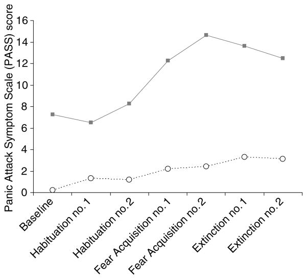
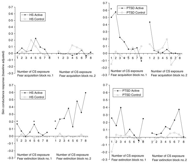
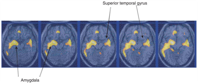
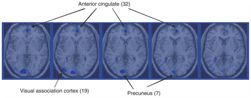
Results: Skin conductance responding to the CS was consistent with the development of conditioned responses with this paradigm. PTSD patients had increased left amygdala activation with fear acquisition, and decreased anterior cingulate function during extinction, relative to controls.

Conclusions: These findings implicate amygdala and anterior cingulate in the acquisition and extinction of fear responses, respectively, in PTSD.


Figures

Fig. 1
Fig. 1
Anxiety symptoms during fear acquisition and extinction as measured with the Panic Attack Symptom Scale (PASS) on the day when electric shock was paired with exposure to the blue square, or conditioned stimulus. Post-traumatic stress disorder (PTSD) patients had elevated anxiety at baseline, and higher levels of anxiety during both acquisition and extinction of fear relative to controls as measured with the PASS. ···· ○····, Control (n=11); —■—, PTSD (n=8).
Fig. 2
Fig. 2
Baseline-corrected skin conductance (SC) during fear acquisition and extinction in healthy subjects and subjects with post-traumatic stress disorder (PTSD). For the active fear acquisition condition subjects were repeatedly exposed (every 10 s) to a paired conditioned stimulus (CS)–unconditioned stimulus (US) consisting of a blue square (CS) appearing on a screen for 4 s with an electric shock (US) starting half a second after the blue square first appeared on the screen. There were two blocks of eight CS–US pairings, each block associated with one PET scan acquisition of brain blood flow. The numbers on the x-axis (1–8) refer to the number of the CS presentation (eight in all for each block). The control for the fear acquisition consisted of an identical procedure except that eight shocks were delivered randomly, i.e. not paired with the CS presentation. The fear acquisition (either active or control blocks) were followed by two blocks of extinction, which consisted of presentation of blue squares for 4 s with a 6-s inter-presentation interval, repeated eight times. The active and control extinction conditions differed only in whether or not they followed the active or control fear acquisition conditions. SC levels are corrected for pre-CS baseline. Asterisks denote times where there were significant elevations in SC in the active compared with the control condition. The number of CS is listed on the x-axis. SC levels were higher for the first block of active fear acquisition versus control in all subjects combined; when the groups were examined separately, increases were only seen in PTSD. Post hoc analyses showed increased SC for the first CS–US pairing in the first block in the PTSD group (* p<0·05). Skin conductance levels were significantly elevated for the first block (but not the second block) in active versus control extinction for PTSD and non-PTSD combined. Differences were significant for non-PTSD only when groups were examined separately. Analysis of individual time-points showed elevations in non-PTSD for the first and fifth presentations of CS in block 1 and the first presentation of CS in block 2.
Fig. 3
Fig. 3
Statistical parametric map overlaid on an MRI template of areas of significantly greater increased blood flow with fear acquisition versus control in post-traumatic stress disorder (PTSD). There was increased blood flow in the amygdala in PTSD. Comparison with non-PTSD showed greater activation in PTSD. Yellow areas correspond to areas of significant activation (Z-score >3·09; p<0·001).
Fig. 4
Fig. 4
Parametric map overlaid on an MRI template of areas of areas of decreased blood flow with extinction in post-traumatic stress disorder (PTSD). Blue areas correspond to areas of significant deactivation (Z-score >3·09; p<0·001). There was decreased blood flow in the medial prefrontal cortex (anterior cingulate, BA 24, 32) in PTSD during extinction relative to control condition in PTSD, not seen in controls.

References

    1. Blake DD, Weathers FW, Nagy LM, Kaloupek DG, Gusman FD, Charney DS. The development of a clinician-administered PTSD scale. Journal of Trauma and Stress. 1995;8:75–90. - PubMed
    1. Blanchard EB, Kolb LC, Gerardi RJ, Ryan P, Pallmeyer TP. Cardiac response to relevant stimuli as an adjunctive tool for diagnosing post-traumatic stress disorder in Vietnam veterans. Behaviour Therapy. 1986;17:592–606.
    1. Bremner JD. Neuroimaging in posttraumatic stress disorder. Psych Annal. 1998;28:445–450.
    1. Bremner JD, Innis RB, Ng CK, et al. PET measurement of cerebral metabolic correlates of yohimbine administration in posttraumatic stress disorder. Archives of General Psychiatry. 1997;54:246–256. - PubMed
    1. Bremner JD, Krystal JH, Putnam F, et al. Measurement of dissociative states with the Clinician Administered Dissociative States Scale (CADSS) Journal of Trauma and Stress. 1998;11:125–136. - PubMed

Publication types