

Detection of asymmetric blotches (asymmetric structureless areas) in dermoscopy images of malignant melanoma using relative color
- PMID: 15998328
- PMCID: PMC3196558
- DOI: 10.1111/j.1600-0846.2005.00117.x
Detection of asymmetric blotches (asymmetric structureless areas) in dermoscopy images of malignant melanoma using relative color
Abstract
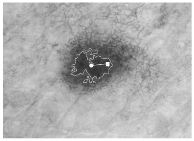
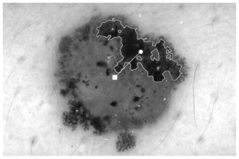
Background: Dermoscopy, also known as dermatoscopy or epiluminescence microscopy (ELM), is a non-invasive, in vivo technique, which permits visualization of features of pigmented melanocytic neoplasms that are not discernable by examination with the naked eye. One prominent feature useful for melanoma detection in dermoscopy images is the asymmetric blotch (asymmetric structureless area).
Method: Using both relative and absolute colors, blotches are detected in this research automatically by using thresholds in the red and green color planes. Several blotch indices are computed, including the scaled distance between the largest blotch centroid and the lesion centroid, ratio of total blotch areas to lesion area, ratio of largest blotch area to lesion area, total number of blotches, size of largest blotch, and irregularity of largest blotch.
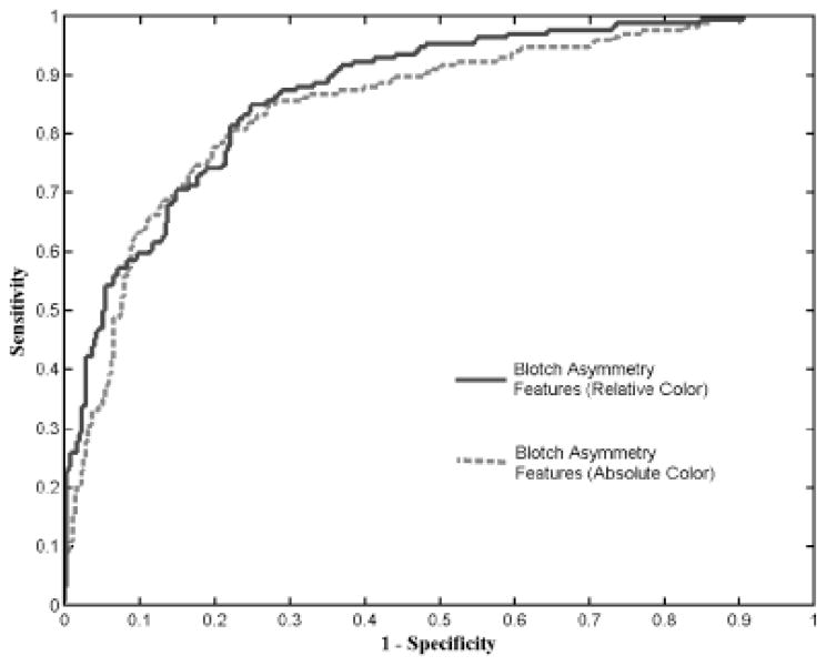
Results: The effectiveness of the absolute and relative color blotch features was examined for melanoma/benign lesion discrimination over a dermoscopy image set containing 165 melanomas (151 invasive melanomas and 14 melanomas in situ) and 347 benign lesions (124 nevocellular nevi without dysplasia and 223 dysplastic nevi) using a leave-one-out neural network approach. Receiver operating characteristic curve results are shown, highlighting the sensitivity and specificity of melanoma detection. Statistical analysis of the blotch features are also presented.
Conclusion: Neural network and statistical analysis showed that the blotch detection method was somewhat more effective using relative color than using absolute color. The relative-color blotch detection method gave a diagnostic accuracy of about 77%.
Figures
References
-
- Jemal A, Tiwari RC, Murray T, et al. Cancer statistics 2004. CA Cancer J Clin. 2004;54:8–29. - PubMed
-
- Nachbar F, Stolz W, Merkle T, et al. The ABCD rule of dermatoscopy. High prospective value in the diagnosis of doubtful melanocytic skin lesions. J Am Acad Dermatol. 1994;30:551–559. - PubMed
-
- Binder M, Schwarz M, Winkler A, et al. Epiluminescence microscopy: a useful tool for the diagnosis of pigmented skin lesions for formally trained dermatologists. Arch Dermatol. 1995;13:286–291. - PubMed
-
- Argenziano G, Fabbrocini G, Carli P, De Giorgi V, Sammarco E, Delfino M. Epiluminescence microscopy for the diagnosis of doubtful melanocytic skin lesions: comparison of the ABCD rule of dermatoscopy and a new 7-point checklist based on pattern analysis. Arch Dermatol. 1998;134:1563–1570. - PubMed
-
- Argenziano G, Soyer HP, Chimenti S, Talamini R, Corona R, Sera F, et al. Dermoscopy of pigmented skin lesions: results of a consensus meeting via the Internet. J Am Acad Dermatol. 2003;48:679–693. - PubMed
Publication types
MeSH terms
Grants and funding
LinkOut - more resources
Full Text Sources
Other Literature Sources
Medical
