Comparative Study
. 2005 Jul 26;102(30):10652-7.
doi: 10.1073/pnas.0504887102. Epub 2005 Jul 5.

Ephrin-B2 ligand is a functional receptor for Hendra virus and Nipah virus

Affiliations
Comparative Study

Ephrin-B2 ligand is a functional receptor for Hendra virus and Nipah virus

Matthew I Bonaparte et al. Proc Natl Acad Sci U S A. .

Abstract

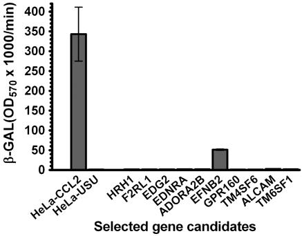
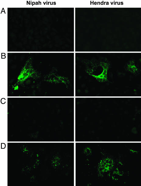
Hendra virus (HeV) and Nipah virus (NiV) belong to the genus Henipavirus of the family Paramyxoviridae and are unique in that they exhibit a broad species tropism and cause fatal disease in both animals and humans. They infect cells through a pH-independent membrane fusion process mediated by their fusion and attachment glycoproteins. Previously, we demonstrated identical cell fusion tropisms for HeV and NiV and the protease-sensitive nature of their unknown cell receptor and identified a human cell line (HeLa-USU) that was nonpermissive for fusion and virus infection. Here, a microarray analysis was performed on the HeLa-USU cells, permissive HeLa-CCL2 cells, and two other permissive human cell lines. From this analysis, we identified a list of genes encoding known and predicted plasma membrane surface-expressed proteins that were highly expressed in all permissive cells and absent from the HeLa-USU cells and rank-ordered them based on their relative levels. Available expression vectors containing the first 10 genes were obtained and individually transfected into HeLa-USU cells. One clone, encoding human ephrin-B2 (EFNB2), was found capable of rendering HeLa-USU cells permissive for HeV- and NiV-mediated cell fusion as well as infection by live virus. A soluble recombinant EFNB2 could potently block fusion and infection and bind soluble recombinant HeV and NiV attachment glycoproteins with high affinity. Together, these data indicate that EFNB2 serves as a functional receptor for both HeV and NiV. The highly conserved nature of EFNB2 in humans and animals is consistent with the broad tropism exhibited by these emerging zoonotic viruses.


Figures

Fig. 1.
Fig. 1.
EFBN2 expression confers HeV fusion permissiveness to HeLa-USU cells. HeLa-CCL2 cells were used as the positive control, and mock-transfected HeLa-USU cells were used as the negative control for cell fusion. Each plasmid (1 μg) was transfected into a single well containing 106 HeLa-USU cells. The cell fusion assay was carried out as detailed in Experimental Procedures. Fusion was obtained only with the EFNB2 gene (human EFNB2). The transfection efficiency was ≈10%, monitored by GFP expression.
Fig. 2.
Fig. 2.
Soluble EFNB2 blocks HeV and NiV fusion in human EFNB2-transfected HeLa-USU cells and HeLa-CCL2 cells. The cells were prepared for fusion as described in Experimental Procedures. (A) HeV-mediated cell fusion with HeLa-CCL2 cells in the presence of anti-EFNB2 Pab or EFNB2/Fc. (B) HeV-mediated cell fusion with HeLa-USU cells expressing human EFNB2 in the presence of anti-EFNB2 Pab or EFNB2/Fc. (C) NiV-mediated cell fusion with HeLa-CCL2 cells in the presence of anti-EFNB2 Pab or EFNB2/Fc. (D) NiV-mediated cell fusion with HeLa-USU cells expressing human EFNB2 in the presence of anti-EFNB2 Pab or EFNB2/Fc. NiV-FC2 (1 μM) was used as a specific inhibitor for NiV- and HeV-mediated fusion in all instances. HeLa-USU cells transfected with G protein-coupled receptor 160 (GPR160) were used as the negative control in B and D.
Fig. 3.
Fig. 3.
Binding of EFNB2 to HeV and NiV G. Two ELISA formats were conducted as detailed in Experimental Procedures. (A) EFNB2/Fc was used as the capture antigen for HeV and NiV sG, detected with anti-sG antisera. (B) HeV and NiV sG were used as capture antigens and detected with either anti-HeV or anti-NiV antisera or EFNB2/Fc. Anti-Tioman virus (TIV) was used as negative control. (C) HeV and NiV coprecipitations. Cell lysates containing myc-tagged metabolically labeled G were precipitated with 9E10 mAb (lanes 1-3) or EFNB2/Fc (lanes 4-6) and protein G (15). Precipitates were separated by SDS/PAGE, and visualized by autoradiography. WR, control vaccimia virus. (D) Interaction between EFNB2/Fc and sG by surface plasmon resonance using a BIACORE 1000. Two surfaces where sG was associated at two different concentrations in two independent experiments were used for test of reproducibility. In all of the experiments EFNB2 was stripped with 10 mM glycine·HCl (pH 2.0), and regeneration did not affect its binding at the same concentration in the next cycle. The arrow indicates the beginning of the dissociation phase.
Fig. 4.
Fig. 4.
Inhibition of HeV (Left) and NiV (Right) by EFNB2/Fc. Vero cells were infected with either HeV or NiV as described in Experimental Procedures. For inhibition, HeV and NiV were preincubated with either 100 or 10 μg/ml EFNB2/Fc for 30 min before addition to the cells. All infections were incubated for 24 h, fixed, and immunofluorescently stained for phosphoprotein before digital microscopy. Images were obtained at an original magnification of ×40.
Fig. 5.
Fig. 5.
HeV (Right) and NiV (Left) infection of EFNB2-transfected HeLa-USU cells. (A) HeLa-USU cells transfected and expressing an irrelevant gene (SH of J virus). (B) HeLa-USU cells expressing human EFNB2. (C) HeLa-USU cells expressing human EFNB2 viruses preincubated with 10 μg/ml EFNB2/Fc before infection. (D) HeLa-CCL2 cells. Infections were conducted as described in Experimental Procedures. After 24 h, cells were fixed and immunofluorescently stained for phosphoprotein before digital microscopy. Images were obtained at an original magnification of ×40.

References

    1. Eaton, B. T. (2001) Microbes Infect. 3, 277-278. - PubMed
    1. Chua, K. B. (2003) J. Clin. Virol. 26, 265-275. - PubMed
    1. Tan, C. T. & Wong, K. T. (2003) Ann. Acad. Med. Singapore 32, 112-117. - PubMed
    1. Anonymous (2005) Commun. Dis. Rep. Wkly. 15, 6.
    1. Anonymous (2004) ProMed-mail, the International Society for Infectious Disease (December 14, 2004, archive no. 20041214.3307). Available at www.promedmail.org. Accessed December 17, 2004.

Publication types

LinkOut - more resources