

Membrane recruitment of NOD2 in intestinal epithelial cells is essential for nuclear factor-{kappa}B activation in muramyl dipeptide recognition
- PMID: 15998797
- PMCID: PMC2171381
- DOI: 10.1083/jcb.200502153
Membrane recruitment of NOD2 in intestinal epithelial cells is essential for nuclear factor-{kappa}B activation in muramyl dipeptide recognition
Abstract
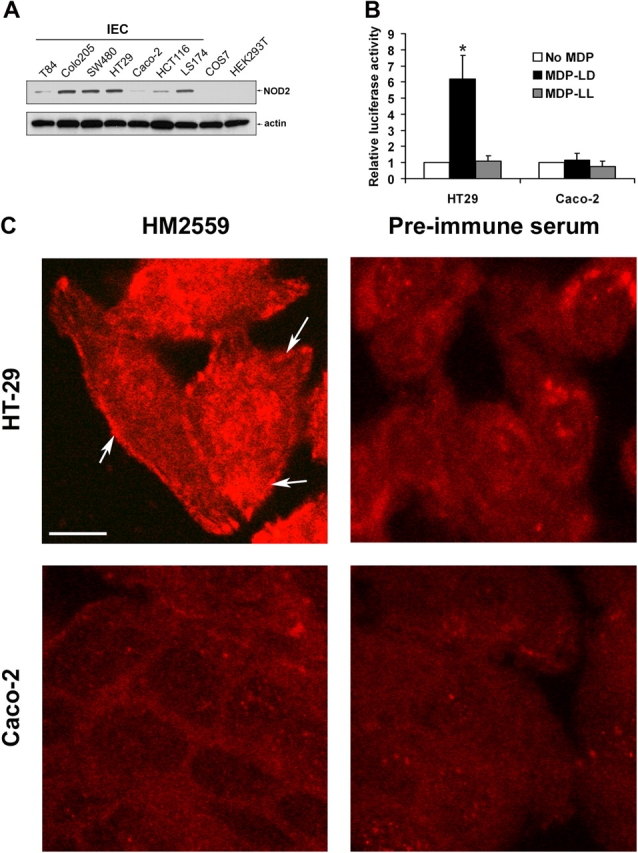
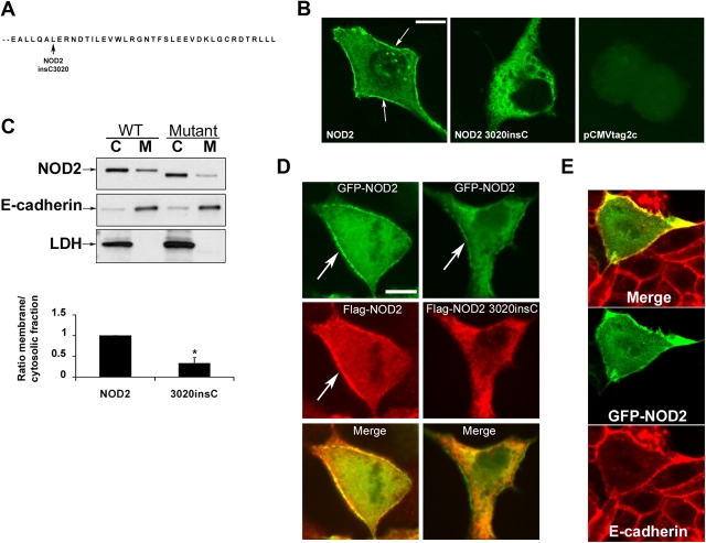
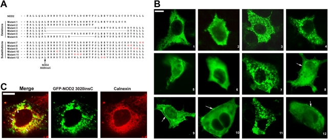
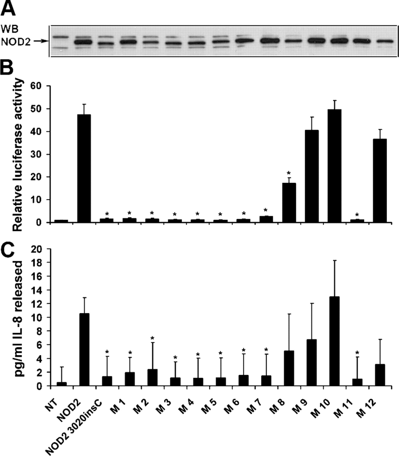
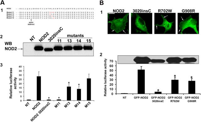
Nucleotide oligomerization domain (NOD) 2 functions as a mammalian cytosolic pathogen recognition molecule, and mutant forms have been genetically linked to Crohn's disease (CD). NOD2 associates with the caspase activation and recruitment domain of RIP-like interacting caspase-like apoptosis regulatory protein kinase (RICK)/RIP2 and activates nuclear factor (NF)-kappaB in epithelial cells and macrophages, whereas NOD2 mutant 3020insC, which is associated with CD, shows an impaired ability to activate NF-kappaB. To gain insight into the molecular mechanisms of NOD2 function, we performed a functional analysis of deletion and substitution NOD2 mutants. NOD2, but not NOD2 3020insC mutant, associated with cell surface membranes of intestinal epithelial cells. Membrane targeting and subsequent NF-kappaB activation are mediated by two leucine residues and a tryptophan-containing motif in the COOH-terminal domain of NOD2. The membrane targeting of NOD2 is required for NF-kappaB activation after the recognition of bacterial muramyl dipeptide in intestinal epithelial cells.
Figures
Similar articles
-
Nod2 mutation in Crohn's disease potentiates NF-kappaB activity and IL-1beta processing.Science. 2005 Feb 4;307(5710):734-8. doi: 10.1126/science.1103685. Science. 2005. PMID: 15692052
-
The NOD2-RICK complex signals from the plasma membrane.J Biol Chem. 2007 May 18;282(20):15197-207. doi: 10.1074/jbc.M606242200. Epub 2007 Mar 13. J Biol Chem. 2007. PMID: 17355968
-
Regulatory regions and critical residues of NOD2 involved in muramyl dipeptide recognition.EMBO J. 2004 Apr 7;23(7):1587-97. doi: 10.1038/sj.emboj.7600175. Epub 2004 Mar 25. EMBO J. 2004. PMID: 15044951 Free PMC article.
-
Nucleotide-binding oligomerization domain containing 2: structure, function, and diseases.Semin Arthritis Rheum. 2013 Aug;43(1):125-30. doi: 10.1016/j.semarthrit.2012.12.005. Epub 2013 Jan 24. Semin Arthritis Rheum. 2013. PMID: 23352252 Review.
-
Inflammatory diseases: is ubiquitinated NEMO at the hub?Curr Biol. 2004 Dec 29;14(24):R1040-2. doi: 10.1016/j.cub.2004.11.040. Curr Biol. 2004. PMID: 15620634 Review.
Cited by
-
NOD2 Polymorphisms and Their Impact on Haematopoietic Stem Cell Transplant Outcome.Bone Marrow Res. 2012;2012:180391. doi: 10.1155/2012/180391. Epub 2012 Oct 18. Bone Marrow Res. 2012. PMID: 23119165 Free PMC article.
-
Cellular stress promotes NOD1/2-dependent inflammation via the endogenous metabolite sphingosine-1-phosphate.EMBO J. 2021 Jul 1;40(13):e106272. doi: 10.15252/embj.2020106272. Epub 2021 May 4. EMBO J. 2021. PMID: 33942347 Free PMC article.
-
Activation and regulation mechanisms of NOD-like receptors based on structural biology.Front Immunol. 2022 Sep 15;13:953530. doi: 10.3389/fimmu.2022.953530. eCollection 2022. Front Immunol. 2022. PMID: 36189327 Free PMC article. Review.
-
Nutrition, IBD and Gut Microbiota: A Review.Nutrients. 2020 Mar 29;12(4):944. doi: 10.3390/nu12040944. Nutrients. 2020. PMID: 32235316 Free PMC article. Review.
-
PGLYRP-1 mediated intracellular peptidoglycan detection promotes mucosal protection.Res Sq [Preprint]. 2024 Oct 14:rs.3.rs-5118704. doi: 10.21203/rs.3.rs-5118704/v1. Res Sq. 2024. Update in: Nat Commun. 2025 Feb 21;16(1):1864. doi: 10.1038/s41467-025-57126-9. PMID: 39483916 Free PMC article. Updated. Preprint.
References
-
- Abbott, D.W., A. Wilkins, J.M. Asara, and L.C. Cantley. 2004. The Crohn's disease protein, NOD2, requires RIP2 in order to induce ubiquitinylation of a novel site on NEMO. Curr. Biol. 14:2217–2227. - PubMed
-
- Barnich, N., T. Hisamatsu, J.E. Aguirre, R. Xavier, H.C. Reinecker, and D.K. Podolsky. 2005. GRIM-19 interacts with NOD2 and serves as down-stream effector of anti-bacterial function in intestinal epithelial cells. J. Biol. Chem. 19:19021–19026. - PubMed
-
- Bertin, J., W.J. Nir, C.M. Fischer, O.V. Tayber, P.R. Errada, J.R. Grant, J.J. Keilty, M.L. Gosselin, K.E. Robison, G.H. Wong, et al. 1999. Human CARD4 protein is a novel CED-4/Apaf-1 cell death family member that activates NF-kappaB. J. Biol. Chem. 274:12955–12958. - PubMed
-
- Bonen, D.K., Y. Ogura, D.L. Nicolae, N. Inohara, L. Saab, T. Tanabe, F.F. Chen, S.J. Foster, R.H. Duerr, S.R. Brant, et al. 2003. Crohn's disease-associated NOD2 variants share a signaling defect in response to lipopolysaccharide and peptidoglycan. Gastroenterology. 124:140–146. - PubMed
Publication types
MeSH terms
Substances
Grants and funding
LinkOut - more resources
Full Text Sources
Other Literature Sources
Molecular Biology Databases
Miscellaneous
