. 2005 Jul 4;170(1):103-13.
doi: 10.1083/jcb.200504008.

Proteomic analysis of a eukaryotic cilium

Affiliations

Proteomic analysis of a eukaryotic cilium

Gregory J Pazour et al. J Cell Biol. .

Abstract

Cilia and flagella are widespread cell organelles that have been highly conserved throughout evolution and play important roles in motility, sensory perception, and the life cycles of eukaryotes ranging from protists to humans. Despite the ubiquity and importance of these organelles, their composition is not well known. Here we use mass spectrometry to identify proteins in purified flagella from the green alga Chlamydomonas reinhardtii. 360 proteins were identified with high confidence, and 292 more with moderate confidence. 97 out of 101 previously known flagellar proteins were found, indicating that this is a very complete dataset. The flagellar proteome is rich in motor and signal transduction components, and contains numerous proteins with homologues associated with diseases such as cystic kidney disease, male sterility, and hydrocephalus in humans and model vertebrates. The flagellum also contains many proteins that are conserved in humans but have not been previously characterized in any organism. The results indicate that flagella are far more complex than previously estimated.


Figures

Figure 1.
Figure 1.
Flagellar structures. Diagram (A) and electron micrograph (B) of a cross section of a motile flagellum (from C. reinhardtii). (C) Central pair of microtubules; (I) inner dynein arm; (IFT) IFT particle; (M) flagellar membrane; (O) outer dynein arm; (R) radial spoke.
Figure 2.
Figure 2.
Flow chart for isolation of flagellar fractions used for MS analyses. (A) Electron micrograph of cross sections of isolated flagella. Most of the flagella have an intact membrane; in these flagella, the matrix is dense and obscures the axonemal microtubules. In a few of the flagella, the membrane has ruptured, releasing the matrix into solution and revealing the microtubules. No other cell organelles are apparent in the flagellar preparation. In the initial analysis (left pathway), isolated flagella from wild-type cells were treated with the detergent Tergitol, which disrupts the flagellar membranes without dissolving them, and releases the flagellar matrix. The Tergitol-insoluble fraction (B) was then collected by centrifugation and analyzed by MS as described in the Materials and methods. For subsequent analyses (right pathway), flagella were isolated from the outer armless mutant oda1 and treated with the detergent Nonidet, which dissolves the membrane and releases the matrix; the preparation was then centrifuged to yield a supernatant containing the “membrane + matrix” fraction and a pellet containing the demembranated axonemes (C). The axonemes were resuspended in 0.6 M KCl and the mixture was centrifuged to yield a supernatant containing the “KCl extract” and a pellet containing the “extracted axonemes” (D). The KCl extraction releases numerous axonemal proteins, including those of the inner dynein arms and the C2 central microtubule, which are missing in the extracted axonemes. The “membrane + matrix,” “KCl extract,” and “extracted axonemes” were then analyzed by MS. Bar, 0.2 μm. (B) Micrograph courtesy of M. Wirschell (Emory University, Atlanta, GA).
Figure 3.
Figure 3.
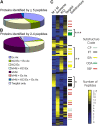
Distribution of proteins in flagellar fractions. (A and B) Distribution of proteins identified by five or more peptides (A) and by two to four peptides (B) in the extracted axoneme (Ex Ax), KCl extract (KCl Ex), and membrane + matrix (M+M) fractions. A protein was assigned to a specific fraction if it was identified by at least twice as many peptides in that fraction as in any other fraction. If the number of peptides in a second or third fraction was more than half that of the first fraction, the protein was assigned to two or all three fractions, as appropriate. Outer dynein arm subunits found exclusively in the Tergitol-insoluble fraction were automatically assigned to the KCl extract fraction. Seven additional proteins were identified by two to four peptides in the Tergitol-insoluble fraction only; these are included in B. (C) Hierarchical cluster diagram. Proteins were clustered based on the distribution of peptides in all four biochemical fractions. Each horizontal row represents one protein. The number of peptides in the particular fraction is represented by a color ranging from yellow (>27 peptides) to blue (0 peptides). In addition, the major known flagellar proteins are color coded under “substructure” with central pair (CP) proteins being marked in blue gray, intraflagellar transport (IFT) proteins in black, inner dynein arm (IDA) proteins in yellow, outer dynein arm (ODA) proteins in green and radial spoke proteins (RSP) in red. Note, for example, the IFT protein cluster (*) where nearly all peptides were found in the M+M fraction, the inner dynein arm cluster (**) where most peptides were most abundant in the KCl extract, and the outer dynein arm cluster (***) where nearly all peptides were found in the Tergitol-insoluble fraction. Only proteins represented by 15 or more peptides were included in the cluster analysis. A detailed version of this figure including gene IDs is available at http://labs.umassmed.edu/chlamyfp/index.php.
Figure 4.
Figure 4.
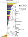
Deflagellation-induced change in transcript levels for 176 predicted flagellar proteins as measured by real-time PCR. (Yellow) Proteins conserved in C. reinhardtii, humans, and A. thaliana (BLAST E ≤1e-10); (blue) proteins conserved in C. reinhardtii and humans but not A. thaliana; (green) C. reinhardtii proteins conserved in A. thaliana but not humans; (red) C. reinhardtii proteins not conserved in humans or A. thaliana. Black dots indicate conserved, uncharacterized proteins. 26 proteins are identified by name or structure. (CP) Central pair protein; (FMG) flagellar membrane glycoprotein; (GAPDH) glyceraldehyde 3-phosphate dehydrogenase; (HSP70) heat-shock protein 70; (IDA) inner dynein arm protein; (IFT) intraflagellar transport protein; (ODA) outer dynein arm protein; (ODA-LC8) dynein light chain 8; (RSP) radial spoke protein; (TUB) tubulin.
Figure 5.
Figure 5.
Conservation of proteins in flagellar proteome. Number of C. reinhardtii (C.r.) flagellar proteins conserved (BLAST E ≤ 1e-10) in H. sapiens (H.s.) and/or A. thaliana (A.t.), out of a total of 652 proteins identified by two or more peptide hits. 292 C. reinhardtii flagellar proteins had no close homologues in either humans or A. thaliana.
Figure 6.
Figure 6.
Enzymes of the glycolytic pathway found in the flagellar proteome. The number of peptides found for each protein is indicated. (HK) Hexokinase; (PGI) phosphoglucose isomerase; (PFK) phosphofructokinase; (GAPDH) glyceraldehyde 3-phosphate dehydrogenase; (PGK) phosphoglycerate kinase; (PGM) phosphoglycerate mutase; (PK) pyruvate kinase; (PC) pyruvate carboxylase; (PEPCK) phosphoenolpyruvate carboxykinase.

Similar articles

Cited by

References

    1. Avidor-Reiss, T., A.M. Maer, E. Koundakjian, A. Polyanovsky, T. Keil, S. Subramaniam, and C.S. Zuker. 2004. Decoding cilia function: defining specialized genes required for compartmentalized cilia biogenesis. Cell. 117:527–539. - PubMed
    1. Bergman, K., U.W. Goodenough, D.A. Goodenough, J. Jawitz, and H. Martin. 1975. Gametic differentiation in Chlamydomonas reinhardtii. II. Flagellar membranes and the agglutination reaction. J. Cell Biol. 67:606–622. - PMC - PubMed
    1. Bloodgood, R.A. 1992. Calcium-regulated phosphorylation of proteins in the membrane-matrix compartment of the Chlamydomonas flagellum. Exp. Cell Res. 198:228–236. - PubMed
    1. Bradley, B.A., J.J. Wagner, and L.M. Quarmby. 2004. Identification and sequence analysis of six new members of the NIMA-related kinase family in Chlamydomonas. J. Eukaryot. Microbiol. 51:66–72. - PubMed
    1. Chae, T.H., S. Kim, K.E. Marz, P.I. Hanson, and C.A. Walsh. 2004. The hyh mutation uncovers roles for alpha Snap in apical protein localization and control of neural cell fate. Nat. Genet. 36:264–270. - PubMed

Publication types

MeSH terms