. 2005 Jul 1;19(13):1532-43.
doi: 10.1101/gad.1318705.

The AIP2 E3 ligase acts as a novel negative regulator of ABA signaling by promoting ABI3 degradation

Affiliations

The AIP2 E3 ligase acts as a novel negative regulator of ABA signaling by promoting ABI3 degradation

Xiuren Zhang et al. Genes Dev. .

Abstract

The phytohormone abscisic acid (ABA) mediates many complex aspects of plant development including seed maturation, dormancy, and germination as well as root growth. The B3-domain transcription factor abscisic acid-insensitive 3 (ABI3) is a central regulator in ABA signaling, but little is known of how this factor is regulated. Here, we show that ABI3 is an unstable protein and that an ABI3-interacting protein (AIP2), which contains a RING motif, can polyubiquitinate ABI3 in vitro. The AIP2 E3 ligase activity is abolished by mutations (C230S; C231S) in the RING motif and the AIP2 (C/S) mutant functions in a dominant-negative manner. AIP2 has a stronger binding affinity for the B2 + B3 domain of ABI3 than the A1 + B1 domain, but only ubiquitinates the latter. In double-transgenic plants, induced AIP2 expression leads to a decrease in ABI3 protein levels. In contrast, ABI3 levels are elevated upon induced expression of the AIP2 RING mutant, which interferes with the endogenous AIP2 E3 activity. An aip2-1-null mutant shows higher ABI3 protein levels compared with wild type after seed stratification, and is hypersensitive to ABA, mimicking the ABI3-overexpression phenotype, whereas AIP2-overexpression plants contain lower levels of ABI3 protein than wild type and are more resistant to ABA, phenocopying abi3. Our results indicate that AIP2 negatively regulates ABA signaling by targeting ABI3 for post-translational destruction.


Figures

Figure 1.
Figure 1.
ABI3 protein is unstable in vivo. (A) ABI3 degradation in vivo can be blocked by MG132. Two-week-old 35S-ABI3–6myc seedlings were transferred to MS liquid medium in the presence of different concentrations of MG132 for 4 h before samples were collected for Western blot analysis. (B) ABI3 protein is short-lived. ABI3 protein levels were analyzed at different times after transfer of 35S-ABI3–6myc plants to MS liquid medium containing cycloheximide (CHX), in the presence or absence of MG132. The molecular mass of ABI3–6myc is ∼150 kDa. A cross-reaction band (asterisk) is shown as a loading control. Relative intensities of ABI3–6myc normalized with respect to the cross-reacting band are given at the bottom.
Figure 2.
Figure 2.
AIP2 is a ubiquitin E3 ligase. (A) Epitope-tagged recombinant AIP2 protein was purified from E. coli extracts. MBP-AIP2–3HA was assayed for E3 activity in the presence or absence of rabbit E1, human E2 (UbcH5b), and 6xHis-ubiquitin (Ub). (B) AIP2 E3 activity is dependent on the integrity of its RING motif. MBP-AIP2–3HA and MBP-AIP2(C/S)–3HA were assayed for self-ubiquitination in the presence of rabbit E1, human E2, and 6xHis-Ub. E2 was omitted in some reactions as control. Western blot analyses were performed by using anti-HA antibody. (MBP) Maltose-binding protein. The arrow indicates either MBP-AIP2–3HA or MBP-AIP2(C/S)–3HA.
Figure 3.
Figure 3.
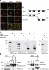
Interaction of AIP2 and ABI3 in vitro and in vivo. (A) Colocalization of AIP2-YFP and ABI3-CFP in the nucleus and the cytosol in N. benthamiana cells. (B) Schematic diagram of full-length and truncated forms of ABI3 (ABI3 ΔC, A1 + B1; ABI3 ΔN, B2 + B3). Numbers refer to the amino acid residues in the wild-type (WT) ABI3 protein. The prey proteins were double tagged with 6xHis and 6myc at N and C termini, respectively, and were purified from E. coli using nitrilotriacetate resin. (C) In vitro pull-down assays of full-length or deletion mutants of ABI3 proteins with three bait proteins (MBP, MBP-SINAT5, and MBP-AIP2). The bait proteins were purified from E. coli using amylose resin. Two micrograms of prey proteins were pulled down with the indicated bait proteins (2 μg each) using amylose resin, and proteins were detected by anti-myc antibody. Another prey protein, ABI5, was used as a negative control. (I) Input amount of prey proteins (asterisks). (D) Coimmunoprecipitation of ABI3 and AIP2 proteins in vivo. Two-week-old seedlings of transgenic seedlings carrying 35-ABI3–6myc/XVE-AIP2(C/S)–3HA were treated overnight with 50 μM MG132 in the absence (lanes 1,4) or presence (lanes 2,3,5) of β-estradiol (25 μM). Total protein extracts were immunoprecipitated (IP) with monoclonal antibody to HA or His. (Left panel) Western blots were analyzed with a polyclonal antibody to myc to detect coimmunoprecipitated ABI3–6myc. (Right panel) In a parallel experiment, IP was done using a polyclonal antibody to myc. Western blots were analyzed with a monoclonal antibody to HA to detect coimmunoprecipitated AIP2–3HA protein. (Input) Crude protein extracts. The arrowhead indicates the cross-reaction with the heavy chain of the protein A-conjugated antibody.
Figure 4.
Figure 4.
Expression patterns of AIP2 and ABI3.(A) Total RNA from different tissues of wild-type (Col-0) plants were analyzed for AIP2 and ABI3 transcripts levels. (B) Four-week-old wild-type (WT) seedlings were transferred to MS medium containing 50 μM ABA or an equal amount of methanol only for the indicated time period (in hours) before RNA extraction. (C) Wild-type seeds in MS medium with or without ABA (5 μM) were stratified at 4°C for 3 d and then transferred to 24 h of continuous light for the indicated days before RNA extraction. Ten micrograms of RNA per lane was used in A, and 5 μg of RNA per lane was used in B and C. The top and middle panels in AC were probed for AIP2 and ABI3 transcripts, respectively. 18S rRNA levels were used as loading controls. (D) Histochemical localization of GUS activity in transgenic plants carrying AIP2-GUS-GFP. GUS expression was detected in germinating seeds at 0 d (panel a), 2 d (panel b), and 4 d (panel c) post-stratification, embryo (panel b), root caps (panels d,g), root vascular tissues (panels e,h), pericycle (panel j), cotyledons (panels c,f,i), shoot apical meristems (panel k), anthers (panel l), pollen sac (panel m), and siliques (panels n,o). Bars: Panels ac,f,ik, 1 mm; panels d,e,g,h, 2 mm; panels mo, 0.5 mm; panel l, 5 mm.
Figure 5.
Figure 5.
ABI3 is a substrate of AIP2 E3 ligase. (A) MBP-AIP2–3HA E3 activity was assayed in the presence or absence of rabbit E1, UbcH5b, 6xHis-ubiquitin, and 6xHis-ABI3–6myc (left panel), 6xHis-ABI3(ΔC)–6myc (middle panel), and 6xHis-ABI3(ΔN)–6myc (right panel). (B) MBP-AIP2 (C/S)–3HA mutant protein has no E3 activity for 6xHis-ABI3–6myc (left panel) and blocks MBP-AIP2–3HA E3 activity by competing for substrates (right panel). Numbers indicate the relative amounts of proteins present in the reaction, where 1 represents 200 ng of MBP, MBP-AIP2–3HA, or MBP-AIP2(C/S)–3HA. (C) Ubiquitination of ABI3 by AIP2 is specific. Reaction mixtures contained 400 ng of MBP-SINAT5 or 400 ng of MBP-COP1. (D) MBP-AIP2 has no E3 activity for 6xHis-HFR1–3HA (Jang et al. 2005) and 6xHis-ABI5–6myc (Lopez-Molina et al. 2003). Western blots were analyzed using polyclonal anti-myc antibody (for full-length and truncated forms of ABI3 and for ABI5) and anti-HA antibody for HFR1. (MBP) Maltose-binding protein. The arrows indicate positions of nonubiquitinated substrates.
Figure 6.
Figure 6.
AIP2 promotes ABI3 protein degradation. (A) The different levels of ABI3 protein levels in wild-type (Col-0), aip2-1 mutant, and AIP2 overexpression lines. (Panel a) T-DNA insertion line (aip2-1) of the AIP2 (At5g20910) gene. Exons are shown as gray boxes and introns as black lines. The predicted translation start (ATG) and stop (TAA) codons are indicated. The 5′- and 3′-untranslated regions are represented by gray lines. The T-DNA insert site in the aip2-1 allele is shown in the upper part. (Panel b) Levels of AIP2 protein in 2-wk seedlings of wild type (Col-0) and aip2-1. (Panels c,d) ABI3 protein levels in wild-type (Col-0), aip2-1 mutant, and 35S-AIP2–3HA/aip2-1 (line #1) seeds. Recombinant 6xHis-AIP2 and 6xHis-ABI3(ΔC)–6myc proteins purified from E. coli were used as positive controls. Tubulin levels were used as loading controls. Seeds were germinated on MS medium supplemented with 3.0 μM ABA for 5 d before Western blot analysis. Endogenous AIP2 and ABI3 were detected with specific polyclonal antibodies generated against the full-length recombinant proteins. The exposure used for panel d was longer than that used for panel c. (B) ABI3 protein levels are reduced after induction of AIP2. Transgenic plants containing 35S-ABI3–6myc and XVE-AIP2–3HA (lines #1, #2, and #3) were treated with and without inducer (25 μM β-estradiol) for 16 h. (C) Induction of AIP2 expression suppresses ABI3 overexpression phenotype. Plants from the same double-transgenic line (line #1) were grown on MS medium for 15 d and then transferred to a fresh medium without (#1 and #2) or with (#3 and #4) β-estradiol (10 μM) for another 15 d. ABI3–6myc and AIP2–3HA levels were determined by Western blots as in B. Bar, 15 mm. (D) ABI3 protein levels are increased by induced expression of AIP2 (C/S) mutant protein. ABI3 and AIP2 protein levels were analyzed by Western blots in three independent transgenic lines (#1, #2, and #3) treated with or without inducer (25 μM β-estradiol). In all assays except A, ABI3 and AIP2 expression levels were detected by anti-myc and anti-HA antibodies, respectively. A cross-reacting band (asterisk) was used to normalize loading. (E) ABA modulates ABI3 stability at the post-translational level by regulating AIP2 expression. Two-week-old seedlings were treated with ABA (50 μM) or an equal amount of methanol before sampling at the indicated times for RNA and Western blot analysis. The cross-reaction bands (arrow and asterisk) in the Western blot were used as loading controls for endogenous AIP2 and ABI3–6myc, respectively. 18S rRNA was used as a loading control in Northern blots.
Figure 7.
Figure 7.
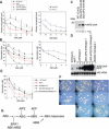
AIP2 is a negative regulator of ABI3 in ABA responses. (A) Five-day-old seedlings germinated in MS were transferred to media containing different concentrations of ABA from 0 to 50 μM, and the length of the primary root was measured 5 d later. The experiment was repeated twice. For each time point, n = 12 seedlings in each independent experiment. (B) The percentage of seeds showing root emergence was scored 6 d post-stratification. Standard error bars represent three independent experiments. Because the germination frequency in MS medium alone is different for each line (lines abi3-1, 35S-AIP2–3HA #8 and #9 have a lower seed viability, ∼80%–95% of wild type), the percent germination without ABA was considered to be 100% and the germination frequency in ABA for these lines was normalized based on this value. The left panels in A and B share the same symbols: Wild-type (Col-0) (square black), aip2-1 (red triangle), 35S-ABI3–6myc (light-gray diamond), 35S-AIP2–3HA line #8 (dark-gray filled circle), and line #9 (dark-gray empty circle). The right panels in A and B show phenotypes of wild type (Ler) (square black) and abi3-1 (blue triangle). n = 50–200. (C) AIP2 protein levels in 35S-AIP2–3HA (#8 and #9) plants in experiments A and B were analyzed by Western blots using anti-HA antibody. Tubulin levels were used as loading controls. (D) Expression of AIP2 and AIP2-HA in aip2-1 mutant and transgenic lines. Two or three independent complemented lines were selected for RNA blots. Five micrograms of total RNA was used in each lane. (E) Germination frequency of aip2-1 mutants and transgenic lines. The percent of seeds showing root emergence was scored 6 d after stratification. Because seeds from different lines for each construct showed similar phenotypes, only a representative line from each construct was graphed for simplicity. The phenotype of pBA002-HA/aip2-1 is the same as that of aip2-1 (data not shown). Wild type (Col-0) (black square), aip2-1 (red triangle), AIP2/aip2-1 (dark-gray filled circle), 35S-AIP2–3HA/aip2-1 (dash line with dark-gray filled cycle), and 35S-AIP2(C/S)–3HA (dashed line with light-gray diamond). Standard error bars represent three replications; n = 100–200. (F) Germinating seeds of the aip2-1 mutant and transgenic lines on MS medium containing 2.5 μM ABA were photographed at 6 d post-stratification. (Panel a) Wild type (Col-0). (Panel b) AIP2/aip2-1 line #1. (Panel c) 35S-AIP2–3HA/aip2-1 line #1. (Panel d) aip2-1. (Panel e) 35S-AIP2(C/S)–3HA/aip2-1 line #1. (Panel f) pBA002–3HA/aip2-1. (Panel g) Wild type (Ler). (Panel h) abi3-1. (G) A working model of the ABA signaling pathway in seed germination. AIP2 promotes ABI3 degradation in ABA signaling. ABI1/ABI2 phosphatases (Leung et al. 1994; Rodriguez et al. 1998) and ERA1 farnesyl transferase (Brady et al. 2003) are negative regulators of ABA signaling and function at or upstream of the ABI3 transcription factor. ABI5, which is negatively regulated by AFP, acts downstream of ABI3 (Finkelstein and Lynch 2000; Lopez-Molina et al. 2002, 2003). ABI4 is another transcriptional factor that acts positively downstream in ABA signaling pathways (Finkelstein et al. 2002).

Similar articles

Cited by

References

    1. Alonso J.M., Stepanova, A.N., Leisse, T.J., Kim, C.J., Chen, H.M., Shinn, P., Stevenson, D.K., Zimmerman, J., Barajas, P., Cheuk, R., et al. 2003. Genome-wide Insertional mutagenesis of Arabidopsis thaliana. Science 301: 653–657. - PubMed
    1. Beaudoin N., Serizet, C., Gosti, F., and Giraudat, J. 2000. Interactions between abscisic acid and ethylene signaling cascades. Plant Cell 12: 1103–1115. - PMC - PubMed
    1. Bechtold N., Ellis, J., and Pelletier, G. 1993. In planta Agrobacterium-mediated gene-transfer by infiltration of adult Arabidopsis thaliana plants. C. R. Acad. Sci. III 316: 1194–1199. - PubMed
    1. Beeckman T. 1994. An easy technique for clearing of histochemically stained plant tissue. Plant Mol. Biol. Rep. 82: 259–266.
    1. Brady S.M., Sarkar, S.F., Bonetta, D., and McCourt., P. 2003. The ABSCISIC ACID INSENSITIVE 3 (ABI3) gene is modulated by farnesylation and is involved in auxin signaling and lateral root development in Arabidopsis. Plant J. 34: 67–75. - PubMed

Publication types