5-HT 1A receptors are reduced in temporal lobe epilepsy after partial-volume correction
- PMID: 16000281
- PMCID: PMC1454475
5-HT 1A receptors are reduced in temporal lobe epilepsy after partial-volume correction
Abstract
Preclinical studies suggest that serotonin 1A receptors (5-HT 1A) play a role in temporal lobe epilepsy (TLE). Previous PET studies reported decreased 5-HT 1A binding ipsilateral to epileptic foci but did not correct for the partial-volume effect (PVE) due to structural atrophy.
Methods: We used PET with 18F-trans-4-fluoro-N-2-[4-(2-methoxyphenyl)piperazin-1-yl]ethyl-N-(2-pyridyl)cyclohexanecarboxamide (18F-FCWAY), a 5-HT 1A receptor antagonist, to study 22 patients with TLE and 10 control subjects. In patients, 18F-FDG scans also were performed. An automated MR-based partial-volume correction (PVC) algorithm was applied. Psychiatric symptoms were assessed with the Beck Depression Inventory Scale.
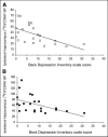
Results: Before PVC, significant (uncorrected P < 0.05) reductions of 18F-FCWAY binding potential (BP) were detected in both mesial and lateral temporal structures, mainly ipsilateral to the seizure focus, in the insula, and in the raphe. Group differences were maximal in ipsilateral mesial temporal regions (corrected P < 0.05). After PVC, differences in mesial, but not lateral, temporal structures and in the insula remained highly significant (corrected P < 0.05). Significant (uncorrected P < 0.05) BP reductions were also detected in TLE patients with normal MR images (n = 6), in mesial temporal structures. After PVC, asymmetries in BP remained significantly greater than for glucose metabolism in hippocampus and parahippocampus. There was a significant inverse relation between the Beck Depression score and the ipsilateral hippocampal BP, both before and after PVC.
Conclusion: Our study shows that in TLE patients, reductions of 5-HT 1A receptor binding in mesial temporal structures and insula are still significant after PVC. In contrast, partial-volume effects may be an important contributor to 5-HT 1A receptor-binding reductions in lateral temporal lobe. Reduction of 5-HT 1A receptors in the ipsilateral hippocampus may contribute to depressive symptoms in TLE patients.
Figures


References
-
- Lu KT, Gean PW. Endogenous serotonin inhibits epileptiform activity in rat hippocampal CA1 neurons via 5-hydroxytryptamine1A receptor activation. Neuroscience. 1998;86:729–737. - PubMed
-
- Clinckers R, Smolders I, Meurs A, Ebinger G, Michotte Y. Anticonvulsant action of hippocampal dopamine and serotonin is independently mediated by D and 5-HT receptors. J Neurochem. 2004;89:834–843. - PubMed
-
- Lang L, Jagoda E, Schmall B, et al. Development of fluorine-18-labeled 5-HT1A antagonists. J Med Chem. 1999;42:1576–1586. - PubMed
-
- Carson RE, Lang L, Fraser C, et al. Human functional imaging with the 5-HT1A ligand [18F]FCWAY [abstract] J Nucl Med. 2002;43(suppl):55P.
-
- Toczek MT, Carson RE, Lang L, et al. PET imaging of 5-HT1A receptor binding in patients with temporal lobe epilepsy. Neurology. 2003;60:749–756. - PubMed
Publication types
MeSH terms
Substances
Grants and funding
LinkOut - more resources
Full Text Sources
Other Literature Sources
Medical
