. 2005 Aug 15;14(16):2323-33.
doi: 10.1093/hmg/ddi235. Epub 2005 Jul 6.

Alms1-disrupted mice recapitulate human Alström syndrome

Affiliations

Alms1-disrupted mice recapitulate human Alström syndrome

G B Collin et al. Hum Mol Genet. .

Abstract

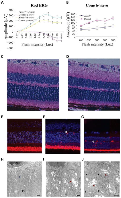
Mutations in the human ALMS1 gene cause Alström syndrome (AS), a progressive disease characterized by neurosensory deficits and by metabolic defects including childhood obesity, hyperinsulinemia and Type 2 diabetes. Other features that are more variable in expressivity include dilated cardiomyopathy, hypertriglyceridemia, hypercholesterolemia, scoliosis, developmental delay and pulmonary and urological dysfunctions. ALMS1 encodes a ubiquitously expressed protein of unknown function. To obtain an animal model in which the etiology of the observed pathologies could be further studied, we generated a mouse model using an Alms1 gene-trapped ES cell line. Alms1-/- mice develop features similar to patients with AS, including obesity, hypogonadism, hyperinsulinemia, retinal dysfunction and late-onset hearing loss. Insulin resistance and increased body weight are apparent between 8 and 12 weeks of age, with hyperglycemia manifesting at approximately 16 weeks of age. In addition, Alms1-/- mice have normal hearing until 8 months of age, after which they display abnormal auditory brainstem responses. Diminished cone ERG b-wave response is observed early, followed by the degeneration of photoreceptor cells. Electron microscopy revealed accumulation of intracellular vesicles in the inner segments of photoreceptors, whereas immunohistochemical analysis showed mislocalization of rhodopsin to the outer nuclear layer. These findings suggest that ALMS1 has a role in intracellular trafficking.


Conflict of interest statement

Conflict of Interest statement. None declared.

Figures

Figure 1
Figure 1
Expression of Alms1 in normal and mutant mice. (A) Insertion site of the gene-trap cassette in intron 13 of the Alms1 gene (BayGenomics, XH152). Locations of PCR primers for genotyping the mice are shown. The presence of the wild-type allele (399 bp) and/or gene-trap allele (447 bp) can be detected with primer pairs WT-F (5′-TGCAATTATGCCTGCATGC-3′) and WT-R (5′-GTTGACCTGGGGAAGGCT-3′) and GT-F (5′-GATCTTATGAGTCACCATATG-3′) and GT-R (5′-GGACTAACAGAAGAACCCGTT-3′), respectively. (B) RT–PCR results of mutant (Alms1−/−) and wild-type (Alms1+/+) eye cDNA. Lanes 1 and 2: cDNA was amplified with oligonucleotides specific for exon 8 and exon 10. Lanes 3 and 4: cDNA was amplified with oligonucleotides specific for exon 13 and exon 16. Lanes 5 and 6: cDNA was amplified with oligonucleotides specific for exon 13 and the gene-trap. Lanes 7 and 8: cDNA was amplified with oligonucleotides specific for exons 22–23, respectively. (Primer locations are shown in Fig. A). By RT–PCR, we observed amplification of exons 22–23, suggesting alternative splicing. (C) Real-time PCR results of mutant (Alms1−/−) and wild-type (Alms1+/+) cDNA. In brain, Alms1 disruption may have resulted in a hypomorphic allele as demonstrated by a low level of exons 13–14 amplicons in the gene-trap mice. By both RT–PCR and real-time PCR, evidence of normal splicing of exons 13–14 was not observed in eye cDNA. (DF) βgal expression in a 7.5-day-old (D), an 8-day-old Alms1+/− heterozygous (E) and a 10.5-day-old Alms1−/− homozygous (F) embryo.
Figure 2
Figure 2
Measures of obesity and diabetes in normal and mutant mice. (A) Truncal obesity in a 20-week-old Alms1−/− mutant mouse. (B–E) Body weight and PG measurements in normal and mutant mice between 4 and 24 weeks of age. Growth curves are shown for female (B) and male (C) homozygous (Alms1−/−; males n = 21–53, females n = 25–56), heterozygous (Alms1+/−; males n = 35–85, females n = 49–102) and wild-type (+/+; males n = 33–48, females n = 27–43) mice, where n is the number of mice weighed per time point. PG levels are shown for female (D) and male (E) Alms1+/+, Alms1+/− and Alms1−/−. Sample sizes for PG were four to 13 animals for each subgroup. Dashed lines represent mean levels indicating transient diabetes. All values are mean ± SEM.
Figure 3
Figure 3
Plasma insulin and lipid measurements in 20-week-old male and female Alms1+/+, Alms1+/− and Alms1−/− mice, respectively. (A) Both male and female mutants exhibit hyperinsulinemia. (B) Plasma concentrations of triglyceride in the mutant and control mice did not differ from controls. (C) Plasma total cholesterols are slightly elevated in mutants (HDL levels are indicated by dotted lines). Sample sizes were five to nine animals for each subgroup. Bar graphs are mean ± SEM.
Figure 4
Figure 4
Light micrographs of affected tissues in Alms1−/− mice; controls (left) and mutant (right). H&E (A, C, E), aldehyde fuchsin (B), and toluidene blue (D). (A) Macro and micro-vesicular lipid deposits (black arrow) accumulate in the liver of a 25-week-old Alms1−/− mouse. (B) Hyperplastic islet is shown in the pancreas of a 24-week-old Alms1−/− mouse. Immunostaining shows an enlarged islet containing insulin-positive beta cells. (C) The testis of the mutant has atrophic seminiferous tubules. Vacuoles (black arrow) are shown surrounding secondary spermatocytes. (D) Sperm heads, stained with toluidene blue in controls (black arrow), are not present in the mutant. (E) Proximal tubules in the mutant kidney are dilated and contain an unidentified flocculent material (black arrow). Scale bars = 50 μm.
Figure 5
Figure 5
Retinal degeneration in Alms1−/− mice. (A and B) Plots of mean amplitudes (± SEM) versus intensity of dark and light adapted electroretinograms of 24-week-old wild-type and mutant mice (Alms1−/−, n = 7; littermate controls, n = 7). Rod and cone b-wave amplitudes are reduced significantly in Alms1−/− mice. H&E stained retinal sections of (C) 24 week wild-type retina and (D) 24-week-old Alms1−/− mutant retinas (10×). (EG) Rhodopsin localization by fluorescent microscopy of retinas tagged with Cy-3 and anti-rhodopsin. (E) 7-week-old wild-type, (F) 7-week-old mutant and (G) 24-week-old mutant (20×). White arrows show the mislocalization of rhodopsin to the outer segments in Alms1−/− mice. (HJ) Ultrastructural analysis of the outer retina. (H) C57BL/6J wild-type adult mouse retina. Typical rough-surfaced endoplasmic reticulum (blue arrowhead) in the inner segments (IS) consists of small, flattened sacs. N indicates the nucleus of ONL. (I and J) Adult Alms1−/− mouse retina. Intracellular vesicles (red arrows) are observed throughout the inner segments, forming stacks resembling the Golgi apparatus. At higher magnification, orientation of the inner segments appears consistently disturbed, often yielding cross-sections (black arrowheads). The white arrows indicate the external limiting membrane and the edge of a photoreceptor nucleus provides orientation. Scale bars (H–J) = 500 nm.
Figure 6
Figure 6
Hearing loss in Alms1−/− mice. At ages between 8 and 12 months, ABR thresholds are higher at all frequencies in Alms1−/− mice (n = 14) when compared with their littermates (n = 11), indicating hearing impairment across the acoustic spectrum.
Figure 7
Figure 7
Ultrastructural analysis of motile and primary cilia in Alms1−/− mice. (A) Cross-section of motile cilia from the nasal epithelium in an Alms1−/− mouse shown by TEM. The microtubule architecture (9+2 arrangement) of the cilia appears normal. (B) Connecting cilia of mutant retina appear intact and are assembled in a typical ‘9+0’ arrangement (data not shown). (C) SEM of primary cilia from renal collecting tubules in an Alms1−/− mouse indicates normal cilia formation.

References

    1. Marshall JD, Ludman MD, Shea SE, Salisbury SR, Willi SM, LaRoche RG, Nishina PM. Genealogy, natural history, and phenotype of Alstrom syndrome in a large Acadian kindred and three additional families. Am J Med Genet. 1997;73:150–161. - PubMed
    1. Michaud JL, Heon E, Guilbert F, Weill J, Puech B, Benson L, Smallhorn JF, Shuman CT, Buncic JR, Levin AV, et al. Natural history of Alstrom syndrome in early childhood: onset with dilated cardiomyopathy. J Pediatr. 1996;128:225–229. - PubMed
    1. Alstrom CH, Hallgren B, Nilsson LB, Asander H. Retinal degeneration combined with obesity, diabetes mellitus and neurogenous deafness: a specific syndrome (not hitherto described) distinct from the Laurence–Moon–Bardet–Biedl syndrome: a clinical, endocrinological and genetic examination based on a large pedigree. Acta Psychiatr Neurol Scand. 1959;34:1–35. - PubMed
    1. Marshall JD, Bronson RT, Collin GB, Nordstrom AD, Maffei P, Paisey RB, Carey C, Macdermott S, Russell-Eggitt I, Shea SE, et al. New Alstrom syndrome phenotypes based on the evaluation of 182 cases. Arch Intern Med. 2005;165:675–683. - PubMed
    1. Collin GB, Marshall JD, Ikeda A, So WV, Russell-Eggitt I, Maffei P, Beck S, Boerkoel CF, Sicolo N, Martin M, et al. Mutations in ALMS1 cause obesity, Type 2 diabetes and neurosensory degeneration in Alstrom syndrome. Nat Genet. 2002;31:74–78. - PubMed

Publication types

MeSH terms