. 2005 Sep;16(9):4350-61.
doi: 10.1091/mbc.e05-04-0304. Epub 2005 Jul 6.

The RNA-binding protein fragile X-related 1 regulates somite formation in Xenopus laevis

Affiliations

The RNA-binding protein fragile X-related 1 regulates somite formation in Xenopus laevis

Marc-Etienne Huot et al. Mol Biol Cell. 2005 Sep.

Abstract

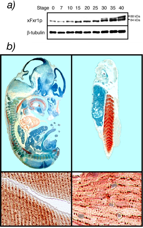
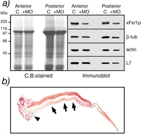
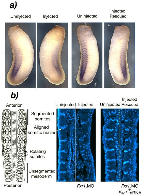
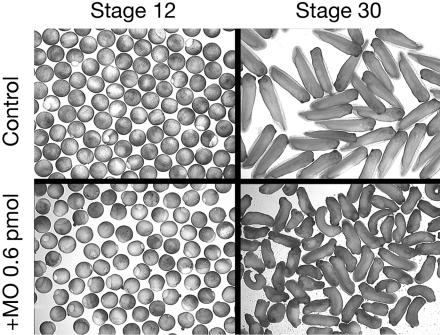
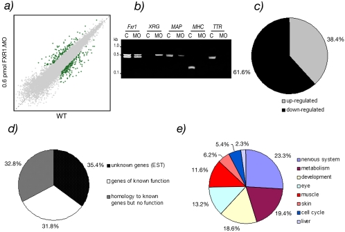
Fragile X-related 1 protein (FXR1P) is a member of a small family of RNA-binding proteins that includes the Fragile X mental retardation 1 protein (FMR1P) and the Fragile X-related 2 protein (FXR2P). These proteins are thought to transport mRNA and to control their translation. While FMR1P is highly expressed in neurons, substantial levels of FXR1P are found in striated muscles and heart, which are devoid of FMRP and FXR2P. However, little is known about the functions of FXR1P. We have isolated cDNAs for Xenopus Fxr1 and found that two specific splice variants are conserved in evolution. Knockdown of xFxr1p in Xenopus had highly muscle-specific effects, normal MyoD expression being disrupted, somitic myotomal cell rotation and segmentation being inhibited, and dermatome formation being abnormal. Consistent with the absence of the long muscle-specific xFxr1p isoform during early somite formation, these effects could be rescued by both the long and short mRNA variants. Microarray analyses showed that xFxr1p depletion affected the expression of 129 known genes of which 50% were implicated in muscle and nervous system formation. These studies shed significant new light on Fxr1p function(s).


Figures

Figure 1.
Figure 1.
Expression of Fxr1p in mouse and frog. Immunoblot analyses of Fxr1p in selected tissues of adult mouse (left) and adult Xenopus (right). Antibody mAb3FX is directed against an epitope common to all Fxr1p isoforms from both mouse and frog, whereas #27-17 is directed against the 27-amino acid pocket coded by the exon 15 that also is present in Xenopus. Fmr1p is detected predominantly in brain extracts from both mouse and frog. Note the cross-reactivity between mAb1C3 and Fxr1p in mouse heart and muscle as described previously (Khandjian et al., 1998; Chemicon International information sheet, MAB2160). H, heart; B, brain; M, muscle; L, liver; and K, kidney.
Figure 2
Figure 2
Fxr1p is conserved in vertebrates. (a) Detection of xFxr1 mRNA variants by RT-PCR in different tissues from mouse and frog by using primers flanking the muscle-specific exon. Northern blots containing RNA isolated from murine and frog tissues (H, heart; B, brain; M, muscle; L, liver; and K, kidney) were hybridized with probes specific for mouse and frog Fxr1. The muscle sequence (exon 15) is found in both mouse and Xenopus. (b) Amino acid sequence of Xenopus Fxr1p (xFxr1p) aligned with human (hFXR1P), mouse (mFxr1p), and zebrafish (zFxr1p) sequences. Conserved amino acids are boxed in black, similar amino acids are in light gray and divergent amino acids are in white. (c) Representation of the mouse (m Iso a-g) and Xenopus (x Iso a and b) Fxr1 mRNA-spliced variants.
Figure 3.
Figure 3.
Nucleotide alignment of human, mouse, and frog 3′-UTR. (a) Short 3′-UTR. (b) Long 3′-UTR. Nucleotides are numbered according to Kirkpatrick et al. (1999) and GenBank accession no. U25165.
Figure 4.
Figure 4.
(a) Developmental profile of xFxr1p accumulation during the first stages of embryogenesis. β-tubulin was used as an internal marker. (b) Immunostained sections of a 12.5 dpc mouse embryo and stage 36 Xenopus embryos reacted with antibody #27-17-specific to the muscle-specific isoforms. Positive signals for Fxr1p were revealed with chromophore AEC (red staining). Counterstaining with hematoxylin (blue).
Figure 5.
Figure 5.
Morphological alterations induced in Xenopus embryos after injection of Fxr1.Morpholino (Fxr1.MO). (a) Lateral and dorsal views of control embryos for the diffusion of the fluorescein tagged Morpholino directed against the human globin (hGlob.MO) injected in one blastomere of two-cell stage embryos. (b) Injected embryos with xFxr1.MO mismatch (mm) as control, and with increasing concentrations of match MO. Number of injected embryos (n =) is on the right of each panel, as well as percentage of penetrance of the depicted phenotypes. The severity of morphological alterations is proportional to the concentration of injected Fxr1.MO (0.3-1.0 pmol). (c) Rescue of the phenotypes is dose-dependent of the injected HA.Fxr1 mRNA.
Figure 6.
Figure 6.
Biochemical evidence that xFxr1p levels decrease after xFxr1.MO treatment. (a) Coomassie-stained proteins after SDS-PAGE analysis of anterior and posterior extracts obtained from one embryo at stage 36 injected with 0.3 pmol of either xFxr1.MOmm (C, control) or xFxr1.MO (+MO). Western blot analysis using antibody mAb3FX for detection of xFxr1p, and antibodies to tubulin, actin, and ribosomal L7 proteins. (b) Partial inactivation of xFxr1correlates with decreased xFxr1p levels in the forming tail. Immunostaining view of a longitudinal section reacted with mAb3FX to detect Fxr1p. Note the reduced levels of xFxr1p in myotomes (arrows) as well as in the head (arrow head) in the injected side of the embryo.
Figure 7.
Figure 7.
Depletion of Fxr1p alters somite formation. (a) In situ hybridization showing the presence of the lateral xMyoD mRNA in the control side, whereas a reduction is observed in the injected side. xMyoD mRNA levels are restored after coinjection with HA.xFr1 mRNA. (b) Left, schematic overview of somite formation in Xenopus. Middle, disruption of nuclear alignment in the injected side of the same embryo depicted in a, as detected by Hoechst staining (right side). Right, somitic nuclei are realigned after rescue with xFxr1 mRNA (same embryo as in a).
Figure 8.
Figure 8.
Injection of 0.6 pmol of xFxr1.MO in both blastomeres at the two-cell stage induces alteration at stage 30. RNA extracted from the control and from the injected embryos were subjected to microarray analyses presented in Figure 7.
Figure 9.
Figure 9.
Microarray analyses of injected embryos with 0.6 pmol of xFxr1.MO versus wild-type embryos. (a) The data set in the squatter plot shows the expression levels of genes on a logarithmic scale as the median value of uninjected (x-axis) and xFxr1.MO injected (y-axis) embryos. The threshold for significancy (p < 0.05) was set at 2.5-fold of change. (b) Examples of validation of RT-PCR analyses of stable (Fxr1), increased (XIRG), and decreased (MAP2, TTR, and MHC) mRNAs in knockdown experiments. (c) Percentage of up- and down-regulated transcripts. (d) Percentage of transcripts of unknown (EST) or known functions, and those presenting homologies to genes not yet described in Xenopus. (e) Tissue distribution of the 129 known transcripts (31.8% in Figure 8d) affected. Note that due to overlapping of these transcripts in different tissues the distribution is only relative and cannot be considered as absolute.

References

    1. Antar, L. N., Afroz, R., Dictenberg, J. B., Carroll, R. C., and Bassell, G. J. (2004). Metabotropic glutamate receptor activation regulates Fragile X mental retardation protein and Fmr1 mRNA localization differentially in dendrites and at synapses. J. Neurosci. 24, 2648-2655. - PMC - PubMed
    1. Bakker, C. E., and The Dutch-Belgian Fragile X Consortium. (1994). Fmr1 knockout mice: a model to study fragile X mental retardation. Cell 78, 23-33. - PubMed
    1. Bakker, C. E., De Diego Otero, Y., Bontekoe, C., Raghoe, P., Luteijn, T., Hoogeveen, A. T., Oostra, B. A., and Willemsen, R. (2000). Immunocytochemical and biochemical characterization of FMRP, FXR1P, and FXR2P in the mouse. Exp. Cell Res. 258, 162-170. - PubMed
    1. Bardoni, B., and Mandel, J.-L. (2002). Advances in understanding of fragile X pathogenesis and FMRP function, and in identification of X linked mental retardation genes. Curr. Opin. Genet. Dev. 12, 284-293. - PubMed
    1. Bisson, N., Islam, N., Poitras, L., Jean, S., Bresnick, A., and Moss, T. (2003). The catalytic domain of xPAK1 is sufficient to induce myosin II dependent in vivo cell fragmentation independently of other apoptotic events. Dev. Biol., 263, 264-281. - PubMed

Publication types

MeSH terms