. 2005 Sep;16(9):4316-28.
doi: 10.1091/mbc.e05-02-0131. Epub 2005 Jul 6.

Src and FAK kinases cooperate to phosphorylate paxillin kinase linker, stimulate its focal adhesion localization, and regulate cell spreading and protrusiveness

Affiliations

Src and FAK kinases cooperate to phosphorylate paxillin kinase linker, stimulate its focal adhesion localization, and regulate cell spreading and protrusiveness

Michael C Brown et al. Mol Biol Cell. 2005 Sep.

Abstract

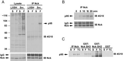
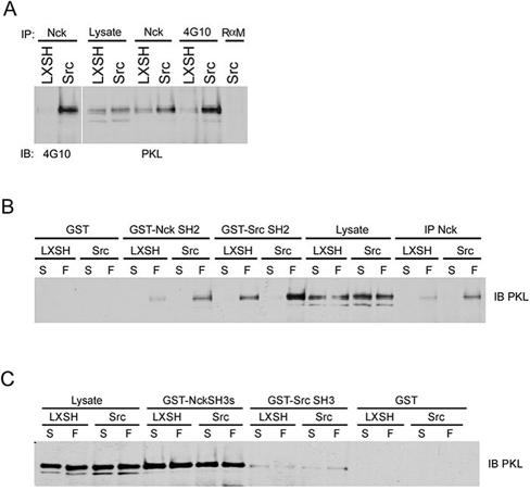
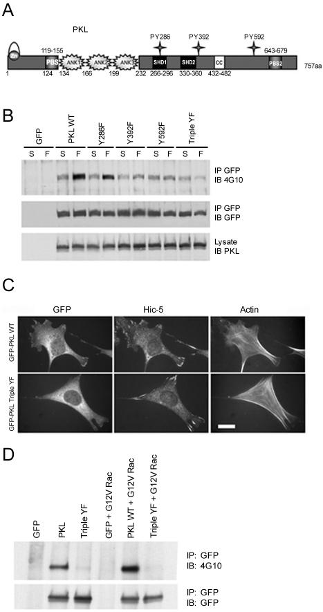
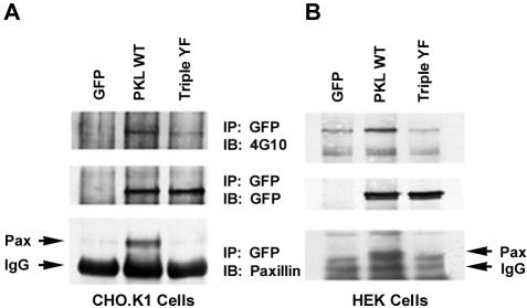
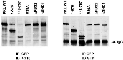
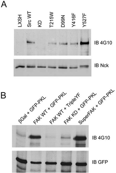
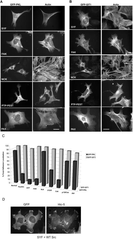
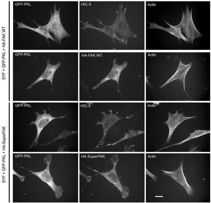
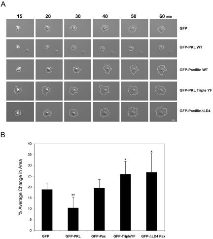
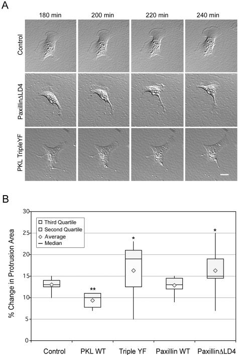
The ArfGAP paxillin kinase linker (PKL)/G protein-coupled receptor kinase-interacting protein (GIT)2 has been implicated in regulating cell spreading and motility through its transient recruitment of the p21-activated kinase (PAK) to focal adhesions. The Nck-PAK-PIX-PKL protein complex is recruited to focal adhesions by paxillin upon integrin engagement and Rac activation. In this report, we identify tyrosine-phosphorylated PKL as a protein that associates with the SH3-SH2 adaptor Nck, in a Src-dependent manner, after cell adhesion to fibronectin. Both cell adhesion and Rac activation stimulated PKL tyrosine phosphorylation. PKL is phosphorylated on tyrosine residues 286/392/592 by Src and/or FAK and these sites are required for PKL localization to focal adhesions and for paxillin binding. The absence of either FAK or Src-family kinases prevents PKL phosphorylation and suppresses localization of PKL but not GIT1 to focal adhesions after Rac activation. Expression of an activated FAK mutant in the absence of Src-family kinases partially restores PKL localization, suggesting that Src activation of FAK is required for PKL phosphorylation and localization. Overexpression of the nonphosphorylated GFP-PKL Triple YF mutant stimulates cell spreading and protrusiveness, similar to overexpression of a paxillin mutant that does not bind PKL, suggesting that failure to recruit PKL to focal adhesions interferes with normal cell spreading and motility.


Figures

Figure 1.
Figure 1.
Identification of a 95-kDa tyrosine phosphorylated protein that associates with Nck in a Src- and adhesion-dependent manner. (A) SYF null cells and SYF + Src replete cells were maintained in suspension or replated on fibronectin-coated dishes followed by anti-Nck immunoprecipitation (IP) and immunoblotting (IB) with anti-phosphotyrosine 4G10 antibody or anti-Nck antibody. A major adhesion induced tyrosine phosphorylated 95-kDa Nck binding partner was identified that was significantly enriched in Src replete versus vector control (LXSH) SYF null cells. (B) SYF + Src cells were plated on 5 μg/ml fibronectin for the indicated times followed by Nck IP and 4G10 anti-phosphotyrosine IB, demonstrating the phos-pho-p95 association with Nck correlates with cell spreading. (C) The tyrosine-phosphorylated p95 protein is precipitated by GST-Nck SH2 and SH3 domains but not GST.
Figure 2.
Figure 2.
Tyrosine phosphorylated PKL binds to Nck and Src. (A) Cultured SYF vector control and Src replete MEFs were lysed followed by anti-Nck or anti-phosphotyrosine 4G10 IP and anti-PKL or 4G10 immunoblotting (IB) revealing PKL is tyrosine phosphorylated and coimmunoprecipitates with Nck. (B) PKL binds to the Nck and Src SH2 domains in an adhesion-dependent manner. SYF or SYF + Src cells were maintained in suspension or replated on fibronectin for 20 min followed by anti-Nck immunoprecipitation (IP), incubation with GST, GST-NckSH2, or GST-SrcSH2 followed by PKL IB. (C) PKL can also bind constitutively to the SH3 domains of Nck and Src.
Figure 3.
Figure 3.
Identification of PKL amino acid sites of phosphorylation and their requirement for PKL localization to focal adhesions. (A) The PKL family of Arf-GAP domain-containing proteins is characterized by an amino-terminal consensus zinc-finger Arf-GAP domain followed by three ankyrin repeat elements. PKL also has two consensus PBSs of which PBS2 has been demonstrated to bind paxillin, two SHDs that may mediate FAK binding, and a coiled-coil (CC) motif that may be involved in dimerization. The potential phosphorylation of three tyrosine residues (286/392/592) that conform with SH2-binding consensus prerequisites was evaluated. (B) SYF + Src cells expressing GFP-PKL tyrosine to phenylalanine point mutants were maintained in suspension or replated on fibronectin for 20 min followed by GFP IP and anti-phosphotyrosine immunoblotting (IB) and reprobing with anti-GFP for loading control. (C) PKL tyrosine phosphorylation is required for localization to focal adhesions. Normal MEFs were plated on fibronectin followed by transfection with GFP-PKL or GFP-PKL TripleYF and constitutively active G12V Rac1. GFP-PKL TripleYF is unable to localize efficiently to focal adhesions (only 25% of transfectants) unlike wild-type GFP-PKL (87% of transfectants). The existence of focal adhesions in GFP-PKL TripleYF transfectants was confirmed by double-labeling with anti-Hic-5. Actin organization was revealed by Alexa350 phalloidin staining. Bar, 20 μm. (D) Active Rac induces tyrosine phosphorylation of GFP-PKL but not the GFP-PKL Triple YF mutant. HEK cells were transfected with GFP-PKL or GFP-PKL Triple YF with or without active G12V Rac. The PKL constructs were precipitated with an anti-GFP antibody and blotted for phosphotyrosine by using 4G10.
Figure 4.
Figure 4.
Tyrosine phosphorylation of PKL is required for binding to paxillin. (A) GFP-PKL or the GFP-PKL Triple YF mutant were expressed in CHO.KI cells and plated on FN for 60 min and then immunoprecipitated with anti-GFP antibody. The precipitates were subjected to anti-phosphotyrosine 4G10, GFP, and paxillin immunoblotting (IB). Only the tyrosine phosphorylated GFP-PKL coprecipitated with paxillin, consistent with a requirement for tyrosine phosphorylation of PKL in focal adhesion targeting via an interaction with Paxillin. (B) The experiment was repeated in HEK293A cells with similar results.
Figure 5.
Figure 5.
PKL targeting to focal adhesions is not required for tyrosine phosphorylation. GFP-PKL constructs were expressed in HEK293A cells followed by replating on fibronection, GFP immunoprecipitation (IP) and anti-phosphotyrosine 4G10 immunoblotting (IB). The nontargeting amino terminus (1-576) and PKLΔPBS2 were phosphorylated demonstrating localization to focal adhesions is not essential for phosphorylation.
Figure 6.
Figure 6.
PKL-Nck association is dependent upon Src and FAK. (A) Nck immunoprecipitates from SYF cells expressing Src mutants were prepared followed by 4G10 anti-phosphotyrosine immunoblotting (IB). PKL phosphotyrosine signal was absent in cells expressing kinase dead Src (KD) but not SH2 (T215W), SH3 (D99N), or autophosphorylation (Y416F) mutants, demonstrating Src kinase activity was required for the association. Notably, cells expressing activated Y527F Src exhibited a stronger phosphotyrosine signal. (B) FAK null MEFs were transfected with GFP-PKL and WT or FAK mutants and plated on fibronectin followed by GFP immunoprecipitation (IP) and 4G10 IB. PKL was not tyrosine phosphorylated in FAK null cells but phosphorylation was rescued upon expression of WT FAK and to a lesser extent with kinase dead FAK (KD). No phosphorylation of GFP-PKL TripleYF was observed upon reexpression of FAK. Cells expressing constitutively active FAK, Super-FAK, had a demonstrably more intense signal.
Figure 7.
Figure 7.
PKL, but not GIT1, requires paxillin, Src and FAK for efficient localization to focal adhesion in MEFs. (A) MEF null cells were plated on fibronectin followed by transfection with GFP-PKL and constitutively active G12VRac1. (A) The capacity of GFP-PKL to localize to focal adhesions was examined by fluorescence microscopy. The actin cytoskeleton was visualized by labeling with rhodamine phalloidin. A significant attenuation in the localization of PKL to focal adhesions in Src, FAK, PTP-PEST, and paxillin but not Nck null cells was observed. (B) The null MEF cells were plated on fibronectin followed by transfection with GFP-GIT1 and G12VRac1 revealing the efficient localization of GIT1 to focal adhesions in all null cells examined. (C) Quantitation of cells, processed as described above, showing focal adhesion localization of GFP-PKL or GFP-GIT1 (expressed as percentage of transfected cells). Unlike PKL, GIT1 retains the capacity to localize efficiently to G12VRac1 focal adhesions irrespective of the loss of Paxillin, Src, or FAK. Bars, 20 μm. (D) Reintroduction of WT Src into the SYF null cells rescues GFP-PKL targeting to focal adhesions.
Figure 8.
Figure 8.
Introduction of SuperFAK(K578/581E) into SYF cells rescues the ability of PKL to localize to focal adhesions. SYF cells were plated on fibronectin, transfected with GFP-PKL, G12VRac1, and either HA-FAK WT or HA-SuperFAK followed by labeling with Al-exa350-phalloidin and Anti-Hic-5 or HA 12CA5 to detect focal adhesion localization of Hic-5 and exogenous FAK, respectively. The capacity of PKL to localize to focal adhesions was then quantified as percentage focal adhesion localization. In the presence of WT-FAK only 12% exhibited GFP-PKL focal adhesion localization, whereas with SuperFAK, 50% of transfectants demonstrated PKL focal adhesion localization. Bar, 10 μm.
Figure 9.
Figure 9.
Expression of GFP-PKL TripleYF increases cell spreading similar to cells expressing paxillin ΔLD4. (A) Normal MEFs were transiently transfected with GFP, GFP-PKL WT, GFP-PKL TripleYF, GFP-Paxillin WT, or GFP-Paxillin ΔLD4 followed by plating on 5 μg/ml fibronectin and acquisition of time-lapse images for 60 min. Both GFP-PKL TripleYF and GFP-Paxillin ΔLD4 increased cell spreading, whereas GFP-PKL WT inhibited cell spreading. GFP or GFP-Paxillin expression was without significant effect. (B) The average change in cell area at 10-min intervals between 20 and 60 min postspreading was calculated. Bars, 10 μm. **p < 0.01, *p < 0.05
Figure 10.
Figure 10.
Expression of GFP-PKL TripleYF increases cell protrusiveness similar to cells expressing paxillin ΔLD4. (A) Normal MEF cells were transfected and plated on 5 μg/ml fibronectin for 180 min followed by acquisition of time-lapse images every 10 min. Both GFP-PKL TripleYF and GFP-Paxillin ΔLD4 exhibited increased cell protrusion and exaggerated retraction fiber formation relative to GFP, suggesting PKL localization is essential for proper integrin-mediated cell function. (B) Changes in cell protrusion areas of at least 10 normal MEF cells transiently transfected and plated as described above were quantitated at 10-min intervals for 1 h as detailed in Materials and Methods, demonstrating both GFP-PKL TripleYF and GFP-Paxillin ΔLD4 exhibited enhanced protrusiveness. Bars, 20 μm. **indicates p < 0.01, *p < 0.05.

References

    1. Andreev, J., Simon, J. P., Sabatini, D. D., Kam, J., Plowman, G., Randazzo, P. A., and Schlessinger, J. (1999). Identification of a new Pyk2 target protein with Arf-GAP activity. Mol. Cell. Biol. 19, 2338-2350. - PMC - PubMed
    1. Angers-Loustau, A., Cote, J. F., Charest, A., Dowbenko, D., Spencer, S., Lasky, L. A., and Tremblay, M. L. (1999). Protein tyrosine phosphatase-PEST regulates focal adhesion disassembly, migration, and cytokinesis in fibroblasts. J. Cell Biol. 144, 1019-1031. - PMC - PubMed
    1. Bagrodia, S., Bailey, D., Lenard, Z., Hart, M., Guan, J. L., Premont, R. T., Taylor, S. J., and Cerione, R. A. (1999). A tyrosine-phosphorylated protein that binds to an important regulatory region on the cool family of p21-activated kinase-binding proteins. J. Biol. Chem. 274, 22393-22400. - PubMed
    1. Bagrodia, S., Taylor, S. J., Jordon, K. A., Van Aelst, L., and Cerione, R. A. (1998). A novel regulator of p21-activated kinases. J. Biol. Chem. 273, 23633-23636. - PubMed
    1. Bladt, F., Aippersbach, E., Gelkop, S., Strasser, G. A., Nash, P., Tafuri, A., Gertler, F. B., and Pawson, T. (2003). The murine Nck SH2/SH3 adaptors are important for the development of mesoderm-derived embryonic structures and for regulating the cellular actin network. Mol. Cell. Biol. 23, 4586-4597. - PMC - PubMed

Publication types

MeSH terms