. 2005 Jul 6;25(27):6322-8.
doi: 10.1523/JNEUROSCI.1405-05.2005.

Cooperative activation of D1 and D2 dopamine receptors enhances a hyperpolarization-activated inward current in layer I interneurons

Affiliations

Cooperative activation of D1 and D2 dopamine receptors enhances a hyperpolarization-activated inward current in layer I interneurons

JianPing Wu et al. J Neurosci. .

Abstract

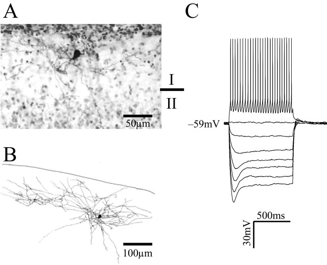
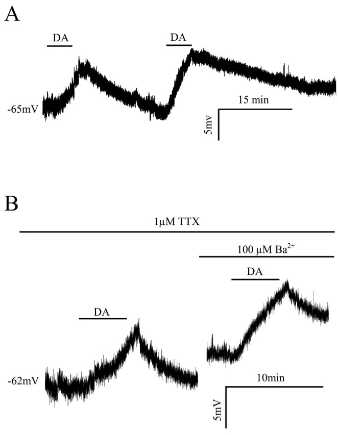
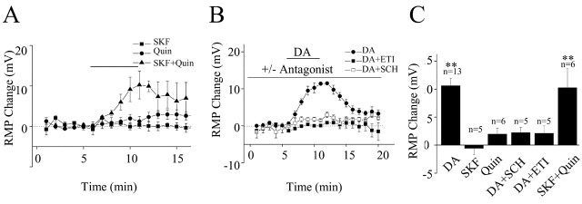
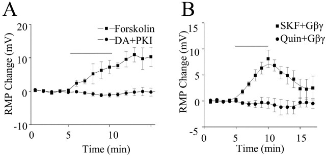
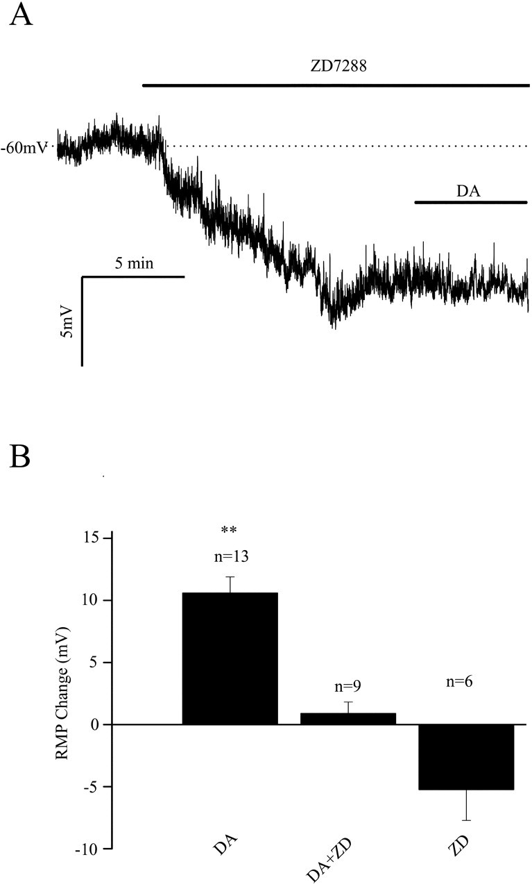
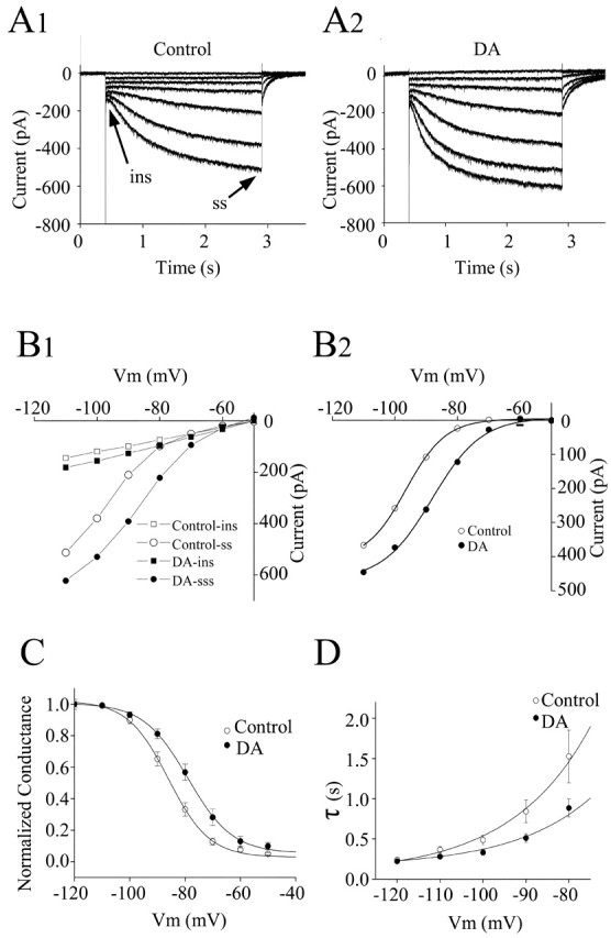
Layer I of the neocortex comprises axonal processes from widespread regions of the brain and a unique population of GABAergic interneurons. Dopamine is known to directly depolarize layer I interneurons, but the underlying mechanism is unclear. Using whole-cell recording techniques in neocortical brain slices, we have examined how dopamine increases excitability of layer I interneurons in postnatal day 7-11 rats. Dopamine (30 microm) caused a 10 mV depolarization of layer I neurons. Paradoxically, neither the D1-like receptor agonist 6-chloro-2,3,4,5-tetrahydro-1-phenyl-1H-3-benzazepine hydrobromide (SKF81297) (1-10 microm) nor the D2-like agonist quinpirole (10 microm) produced a significant depolarization. Depolarization was observed when SKF81297 and quinpirole were coapplied. When G-protein betagamma subunits were included in the recording pipette, D1 but not D2 agonists depolarized layer I neurons. Bath application of 4-ethylphenylamino-1,2-dimethyl-6-methylaminopyrimidinium chloride, a specific blocker of inwardly rectifying hyperpolarization-activated current (Ih) channels, hyperpolarized the neurons and occluded the action of dopamine. Voltage-clamp analysis demonstrated that dopamine increased the amplitude and shifted the voltage dependence of activation of Ih. These results indicate that Ih contributes to the resting potential of layer I interneurons and is subject to modulation by dopamine.


Figures

Figure 1.
Figure 1.
Identification of layer I interneurons in the rat prefrontal cortex. A, Photomicrograph of a biocytin-labeled layer I interneuron. B, Camera lucida reconstruction of the neuron from A. This interneuron had an extensive dendritic tree restricted to the layer I and four axon collaterals that descended to layer II/III. C, Current-clamp recording from the interneuron shown in A. Injection of a depolarizing current elicited a train of action potentials that showed no accommodation. Action potential durations were brief (base duration, ∼2 ms) and were followed by a fast afterhyperpolarization. Hyperpolarizing current pulses induced a hyperpolarization that reached a peak and sagged back toward rest. Such responses are a hallmark of hyperpolarization-activated, nonspecific cation channels.
Figure 2.
Figure 2.
Dopamine depolarizes layer I interneurons in the prefrontal cortex. A, Typical whole-cell current-clamp recording showing that dopamine (50 μm) reversibly depolarizes layer I interneurons. Depolarization response to dopamine recovered after a 10-20 min wash. Reapplication of dopamine induced a similar response in this cell. B, In the presence of 1 μm TTX, dopamine (50 μm) depolarized layer I interneurons to a similar extent as seen in untreated conditions, indicating the depolarization was mediated by a direct effect of dopamine on the recorded cell and is independent of synaptic transmission. In the same interneuron after recovery from the first dopamine application, bath applying 100 μm Ba2+ induced an ∼5 mV depolarization because of the inhibition of a Ba2+-sensitive inward rectifier potassium current. Application of dopamine in the presence of Ba2+ elicited a membrane depolarization comparable with the control condition, suggesting dopamine depolarization of layer I interneurons does not involve modulation of a Ba2+-sensitive inward rectifier current. DA, Dopamine.
Figure 3.
Figure 3.
Synergistic activation of D1- and D2-like receptors is required for dopamine-induced membrane depolarization of layer I neurons. A, Summary time course plot of the effect of the specific D1 and D2 agonists SKF81297 and quinpirole, respectively. Neither SKF81297 (1 μm) nor quinpirole (10 μm) depolarized layer I interneurons. Coapplication of SKF81297 (1 μm) and quinpirole (10 μm) elicited depolarizations similar to that produced by dopamine. B, Summary time course plot of the effect of pretreatment with the selective D1-like receptor antagonist SCH23390 (10 μm) or the D2-like receptor antagonist eticlopride (1 μm). Both antagonists blocked the dopamine-induced membrane depolarization. C, Bar graph of averaged membrane potential changes induced by dopamine, SKF81297, quinpirole, dopamine in the presence of SCH23390 or eticlopride, and SKF81297 plus quinpirole. RMP, Resting membrane potential; SKF, SKF81297; Quin, quinpirole; SCH, SCH23390; ETI, eticlopride; DA, dopamine. **p < 0.01.
Figure 4.
Figure 4.
Requirement of cAMP-dependent PKA activation for dopamine-induced membrane depolarization. A, Forskolin (10 μm), a potent nonspecific adenylyl cyclase activator, produced a significant membrane depolarization. The effect of forskolin was persistent and difficult to reverse after washing. The addition of 1 μm PKA inhibitory peptide (PKI) in the recording pipette blocked the dopamine (DA)-induced depolarization. In the PKI experiments, dopamine was applied at least 15 min after achieving the whole-cell configuration to allow diffusion of the peptide into the cell. The perfusion of PKI itself did not induce significant membrane potential changes. B, Effect of including purified bovine brain G-protein βγ (Gβγ; 20 nm) in the recording pipette. The D1-like receptor agonist SKF81297 (SKF; 1 μm) produced a significant depolarization when Gβγ was included in the recording pipette; the D2-like antagonist quinpirole (Quin; 10 μm) did not. These results strongly suggest that synergistic activation of D1 and D2 receptors by dopamine produces a depolarization of layer I interneurons via a mechanism involving Gβγ and cAMP signaling. RMP, Resting membrane potential.
Figure 5.
Figure 5.
Effects of ZD7288, a specific Ih blocker, on dopamine-induced membrane depolarizations. A, A typical whole-cell current-clamp recording shows the effects of Ih inhibition. When 30 μm ZD7288 was bath applied, a reproducible hyperpolarization was observed, indicating that Ih contributes to the resting membrane potential of these cells. Application of dopamine in the presence of ZD7288 did not produce a depolarization. B, Summary plot of ZD7288 effects. Ih inhibition hyperpolarized interneurons and occluded the depolarizing effect of dopamine. DA, Dopamine; RMP, resting membrane potential; ZD, ZD7288. p < 0.01.
Figure 6.
Figure 6.
Voltage-clamp analysis of the effects of dopamine on Ih activation. A1, Voltage-clamp recordings of Ih. The neuron was voltage clamped at -50 mV, and a series of hyperpolarizing voltage commands from -110 to -50 in 10 mV steps were applied. A time- and voltage-dependent inward current was activated by steps more negative than -70 mV. The instantaneous current (arrow) was measured just after the decay of the capacitive transient, and the steady-state current (arrow) was measured near the end of each voltage commend. A2, In the presence of 30 μm dopamine, Ih amplitude was enhanced and there was a depolarizing shift in activation threshold (-60 mV). After dopamine application, the holding current increased ∼65 pA corresponding to the depolarization seen in current-clamp recordings. The holding current change has been subtracted. B1, Current-voltage curve showing the instantaneous (▪ and □) and steady-state (○ and •) currents as a function of the commend voltage in control and in the presence of 30 μm dopamine. Dopamine increased the steady-state current at all voltages negative to -60 mV and had little effect on the instantaneous current. B2, Plot of the Ih amplitude measured as the difference between steady-state and instantaneous current under control conditions (•) and in the presence of dopamine (○). C, Dopamine caused a depolarizing shift in the voltage dependence of Ih activation. Ih amplitude from 19 interneurons was converted to conductance (g) with the equation g = Ih/(E - Erev), where E is the command voltage and Erev is the reversal potential of Ih; the value of Erev is -27 mV by calculating the tail current. The conductance g for each cell was normalized by Gmax (g/Gmax); G was taken to be the value of g at -110 mV, which was assumed to reach the maximum activation. The normalized conductance (g/Gmax ± SE) in control (▪) and dopamine (•) were plotted as a function of voltage. The data were fitted with the Boltzman equation [g/G = 1/(1 + e(v-v1/2)/K)]; dopamine shifted the curve to the right by changing the V1/2 from -86.0 ± 0.6 mV to -78.9 ± 0.4 mV (n = 19; p < 0.01). D, Plot of the rate of Ih activation as a function of voltage. ins, Instantaneous current; ss, steady-state current; DA, dopamine; Vm, voltage.

References

    1. Berger B (1992) Dopaminergic innervation of the frontal cerebral cortex: evolutionary trends and functional implications. In: Advances in neurology (Chauvel P, Delgado-Escueta AV, Haalgren E, Bancaud J, eds), pp 525-544. New York: Raven. - PubMed
    1. Bernardi G, Cherubini E, Marciani MG, Mercuri N, Stanzione P (1982) Responses of intracellularly recorded cortical neurons to the iontophoretic application of dopamine. Brain Res 245: 267-274. - PubMed
    1. Bradford R, Parnavelas JG, Lieberman AR (1978) Neurons in layer I of the developing occipital cortex of the rat. J Comp Neurol 176: 121-132. - PubMed
    1. Chen S, Wang J, Siegelbaum SA (2001) Properties of hyperpolarization-activated pacemaker current defined by coassembly of HCN1 and HCN2 subunits and basal modulation by cyclic nucleotide. J Gen Physiol 117: 491-504. - PMC - PubMed
    1. Christophe E, Roebuck A, Staiger JF, Lavery DJ, Charpak S, Audinat E (2002) Two types of nicotinic receptors mediate an excitation of neocortical layer I interneurons. J Neurophysiol 88: 1318-1327. - PubMed

Publication types

MeSH terms

LinkOut - more resources