Comparative Study
. 2005 Jul 6;25(27):6401-8.
doi: 10.1523/JNEUROSCI.1563-05.2005.

A highly specific inhibitor of matrix metalloproteinase-9 rescues laminin from proteolysis and neurons from apoptosis in transient focal cerebral ischemia

Affiliations
Comparative Study

A highly specific inhibitor of matrix metalloproteinase-9 rescues laminin from proteolysis and neurons from apoptosis in transient focal cerebral ischemia

Zezong Gu et al. J Neurosci. .

Erratum in

  • J Neurosci. 2005 Nov 9;25(45):10576

Abstract

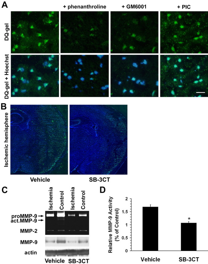
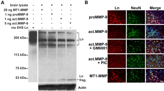
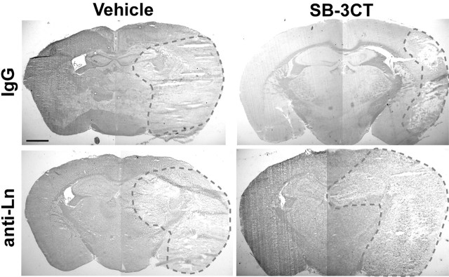
Neuronal cell death occurs during many neurodegenerative disorders and stroke. The aberrant, excessive activity of matrix metalloproteinases (MMPs), especially MMP-9, contributes directly to neuron apoptosis and brain damage (Rosenberg et al., 1996; Asahi et al., 2001; Gu et al., 2002; Horstmann et al., 2003). We determined that MMP-9 degrades the extracellular matrix protein laminin and that this degradation induces neuronal apoptosis in a transient focal cerebral ischemia model in mice. We also determined that the highly specific thiirane gelatinase inhibitor SB-3CT blocks MMP-9 activity, including MMP-9-mediated laminin cleavage, thus rescuing neurons from apoptosis. We conclude that MMP-9 is a highly promising drug target and that SB-3CT derivatives have significant therapeutic potential in stroke patients.


Figures

Figure 1.
Figure 1.
Thiirane inhibitor SB-3CT protects against brain damage and ameliorates neurological outcome after transient focal cerebral ischemia in mice. A, Laser Doppler flowmetry of rCBF. rCBF was measured over the ischemic core of the right MCA region, and the preischemic rCBF was assigned a value of 100% at baseline. For each mouse, rCBF decreased to < 25% of the baseline value during a 2 h period of ischemia and recovered to >50% of baseline during reperfusion. There were no significant differences in rCBF between vehicle-treated controls (solid squares; n = 11) and SB-3CT-treated animals (solid triangles represent mean ± SEM; n = 13). B, Representative TTC staining of stroke in mouse-brain sections after SB-3CT treatment versus vehicle-treated control (Vehicle). SB-3CT (25 mg/kg body weight per treatment) was administrated intraperitoneally as a suspension in a vehicle solution (10% DMSO in saline). SB-3CT was administered in four groups plus parallel vehicle-treated control groups: a preischemic group treated 0.5 h before insult (-0.5 h) and groups treated 2, 6, or 10 h after ischemia (labeled +2, +6, and +10 h; see Materials and Methods). Coronal sections, 1 mm in thickness, were prepared and stained with TTC. C, Quantification of infarct volume by TTC staining. Infarct volumes were determined 24 h after reperfusion. SB-3CT decreased infarct volume compared with vehicle-treated controls (Ctrl). Data represent mean ± SEM. The numbers of animals in each group are as follows: Ctrl, n = 19; SB-3CT, n = 23. *p < 0.001 by ANOVA. D, Neurological behavioral score (see Materials and Methods) 24 h after MCA occlusion/reperfusion. Treatment with SB-3CT (n = 31) significantly improved neurological function compared with vehicle-treated controls (n = 22); *p < 0.02 by ANOVA. Error bars represent SEM.
Figure 2.
Figure 2.
Thiirane inhibitor SB-3CT inhibits MMP-9 activity and consequent increased expression of MMP-9 in the ischemic mouse brain after transient middle cerebral artery occlusion. A, In situ zymography with the MMP fluorogenic substrate DQ-gel (green in top panels) merged with nuclear DNA staining by Hoechst dye (blue plus green in bottom panels). The broad-spectrum MMP inhibitors 1,10-phenanthroline and GM6001, but not a non-MMP PIC, abrogated MMP gelatinolytic activity in the ischemic cortex after MCA occlusion/reperfusion. Scale bar, 25 μm. B, SB-3CT significantly reduced MMP gelatinolytic activity in the ischemic region compared with the vehicle-treated control, as demonstrated by deconvolution microscopy. C, Gelatin zymography and Western blotting reveal upregulation of proMMP-9 (92 kDa) and activation of MMP-9 (act.MMP-9) in the ischemic brain compared with the contralateral hemisphere. In contrast, MMP-2 was not affected. SB-3CT attenuated the increase in proMMP-9 and act.MMP-9. Actin was used as a loading control. D, Quantification of relative MMP-9 activity by densitometry of gelatin zymography. Vehicle, n = 8; SB-3CT, n = 6; *p < 0.0001. Error bars represent SEM.
Figure 3.
Figure 3.
Increased MMP gelatinolytic activity is spatially associated with neuronal laminin in the ischemic cortex of mouse brains after transient middle cerebral artery occlusion. Left panels, Double-immunofluorescent staining revealed two types of morphology, representing Ln (red) on elongated microvascular structures and on the neuronal surface (neurons labeled with the neuron-specific marker anti-NeuN; green). Top left, Inset, Immunolabeling of the α-5 subunit of laminin-10 (Lnα5), which is specific to neurons (Indyk et al., 2003). Right panels, Increased MMP gelatinolytic activity (DQ-gel; green) colocalized with laminin detected by immunostaining with a pan-Ln antibody (red) in the ischemic cortex 2 h after reperfusion. Bottom panels, Merged images counterstained with Hoechst dye to visualize nuclei (blue). Scale bar, 25 μm.
Figure 4.
Figure 4.
Exogenous MMP-9 degrades lamininin the extracellular matrix protein of mouse brain. A, Western blot with a pan-Ln polyclonal antibody reveals degradation of laminin (especially the 360 and 170 kDa subunits) to a 51 kDa fragment (frag.) in brain lysates treated with activated MMP-9 but not with latent proMMP-9 or catalytic MT1-MMP (50 μg of total protein per lane). Purified mouse Engelbreth-Holm-Swarm laminin (ms EHS Ln) served as a molecular marker for laminin immunoblotting. The membrane was reblotted with anti-actin antibody to ensure equal protein loading in each lane. B, Ex vivo degradation of neuronal laminin by exogenous MMP-9 in mouse-brain sections. Double immunolabeling of laminin by pan-Ln polyclonal antibody (Ln; red) and neurons with NeuN antibody (green) reveals that activated MMP-9 degraded neuronal laminin. A broad-spectrum MMP inhibitor, GM6001, significantly reduced laminin degradation, whereas a non-MMP PIC did not. Latent proMMP-9 or catalytic MT1-MMP could not degrade neuronal laminin. Merged images were counterstained with Hoechst dye to visualize nuclei (blue). Scale bar, 25 μm.
Figure 5.
Figure 5.
NO-activated MMP-9 leads to laminin degradation in the ischemic cortex after MCA occlusion/reperfusion. A, Laminin immunoreactivity (red) and Hoechst DNA stain (blue). Deconvolution microscopy revealed that laminin immunoreactivity was significantly reduced in the ischemic cortex of wild-type mice (top right) compared with the contralateral nonischemic control hemisphere (top left). Laminin degradation in the ischemic cortex was attenuated after MCA occlusion/reperfusion in either wild-type mice treated with the specific nNOS inhibitor 3-bromo-7-nitroindazole (3br7NI; bottom left) or in nNOS KO mice (bottom right). Scale bar, 25 μm. B, Quantification of Ln-positive cells in cortex was determined 24 h after reperfusion. Data represent mean ± SEM on 600-1000 cells counted from each brain section (n = 5 in each group; *p < 0.001 compared with control group and #p < 0.001 compared with ischemic group by ANOVA). Error bars represent SEM.
Figure 6.
Figure 6.
Time course of laminin degradation and apoptotic cell death in the ischemic cortex after transient MCAO/R in mice. A-C, In situ zymography reveals that increased MMP gelatinolytic activity (A; green) is associated with apoptotic cell death detected by TUNEL (B; red). Merged images were counterstained with Hoechst dye to visualize nuclei (C; blue). D-F, After 2 h focal cerebral ischemia plus 3 h reperfusion (E) or 24 h reperfusion (F), animals were killed, and the brains were processed for immunohistochemistry. Coronal brain sections were stained for laminin immunoreactivity (red) and nuclear DNA staining with Hoechst dye (blue). D, Immunostaining with anti-pan-Ln antibody (red; indicated by arrowheads) was decreased in the ischemic cortex as early as 3 h after reperfusion compared with the control contralateral cortex. The remaining laminin was mostly associated with elongated microvascular structures rather than neurons. Modest neuronal cell death occurred at 3 h but more massive death at 24 h (arrows), as evidenced by condensed nuclei. G, Quantification of Ln-positive and apoptotic cells in the cortex 24 h after reperfusion. Data represent mean ± SEM on 600-1000 cells counted from each brain section; n = 5 in each group; *p < 0.001 by ANOVA compared with Ln-positive cells in the control contralateral hemisphere (Ctrl; filled bar), and #p < 0.001 compared with apoptotic cells in the control contralateral hemisphere (Ctrl; open bar). H, I, Coronal brain sections were stained for laminin immunoreactivity (green) and TUNEL (red) to demonstrate the reduction in laminin and increase in apoptosis in the ischemic cortex (I) compared with the contralateral control cortex (H). Brain sections were counterstained with Hoechst dye to show nuclei (blue). Together, the data in this figure suggest that MMP-induced laminin degradation occurs before neuronal apoptotic-like cell death. Scale bar, 25 μm. Error bars represent SEM.
Figure 7.
Figure 7.
SB-3CT attenuates laminin degradation in the ischemic hemisphere after MCAO/R. Western blot demonstrates laminin proteolysis (especially of the 360 and 170 kDa subunits) to a 51 kDa fragment in the ischemic brain (arrowhead at bottom of gel), whereas treatment with SB-3CT decreased laminin degradation after transient MCAO/R. The ∼60 kDa fragment may represent an additional proteolytic derivative of the γ subunit (bottom molecular band) lacking NH2-terminal residues, as reported previously (Giannelli et al., 1997). The membrane was reprobed with anti-actin antibody to ensure equal loading.
Figure 8.
Figure 8.
Disruption of laminin-cell surface interactions increases sensitivity to ischemic death. Mouse brains were infused with normal rabbit serum (IgG) or with a neutralizing antibody to pan-laminin (anti-Ln) in 1% BSA/PBS for 2 d before MCAO/R plus SB-3CT treatment or vehicle only. Brain sections were stained with cresyl violet and acid fuchsin. The dashed red line encircles the area of cell death. Pan-laminin antibody increased cell death in the MCAO/R mouse model despite SB-3CT treatment. This finding is consistent with the notion that the action of anti-laminin antibody is downstream to MMP-9 activation. Scale bar, 1 mm.

References

    1. Asahi M, Asahi K, Jung JC, del Zoppo GJ, Fini ME, Lo EH (2000) Role for matrix metalloproteinase 9 after focal cerebral ischemia: effects of gene knockout and enzyme inhibition with BB-94. J Cereb Blood Flow Metab 20: 1681-1689. - PubMed
    1. Asahi M, Wang X, Mori T, Sumii T, Jung JC, Moskowitz MA, Fini ME, Lo EH (2001) Effects of matrix metalloproteinase-9 gene knock-out on the proteolysis of blood-brain barrier and white matter components after cerebral ischemia. J Neurosci 21: 7724-7732. - PMC - PubMed
    1. Brown S, Bernardo MM, Li ZH, Kotra LP, Tanaka Y, Fridman R, Mobashery S (2000) Potent and selective mechanism-based inhibition of gelatinases. J Am Chem Soc 122: 6799-6800.
    1. Castellanos M, Leira R, Serena J, Pumar JM, Lizasoain I, Castillo J, Davalos A (2003) Plasma metalloproteinase-9 concentration predicts hemorrhagic transformation in acute ischemic stroke. Stroke 34: 40-46. - PubMed
    1. Chen ZL, Strickland S (1997) Neuronal death in the hippocampus is promoted by plasmin-catalyzed degradation of laminin. Cell 91: 917-925. - PubMed

Publication types

MeSH terms