. 2005 Jul;71(7):3786-96.
doi: 10.1128/AEM.71.7.3786-3796.2005.

Suppression of damping-off disease in host plants by the rhizoplane bacterium Lysobacter sp. strain SB-K88 is linked to plant colonization and antibiosis against soilborne Peronosporomycetes

Affiliations

Suppression of damping-off disease in host plants by the rhizoplane bacterium Lysobacter sp. strain SB-K88 is linked to plant colonization and antibiosis against soilborne Peronosporomycetes

Md Tofazzal Islam et al. Appl Environ Microbiol. 2005 Jul.

Abstract

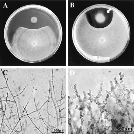
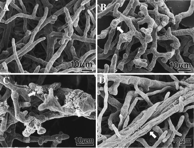
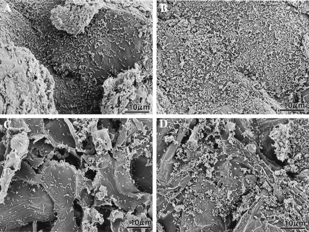
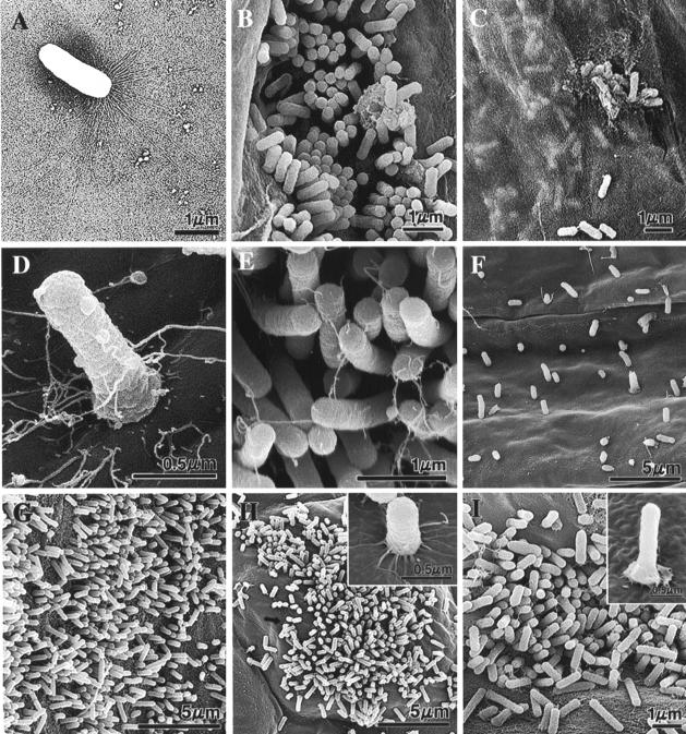
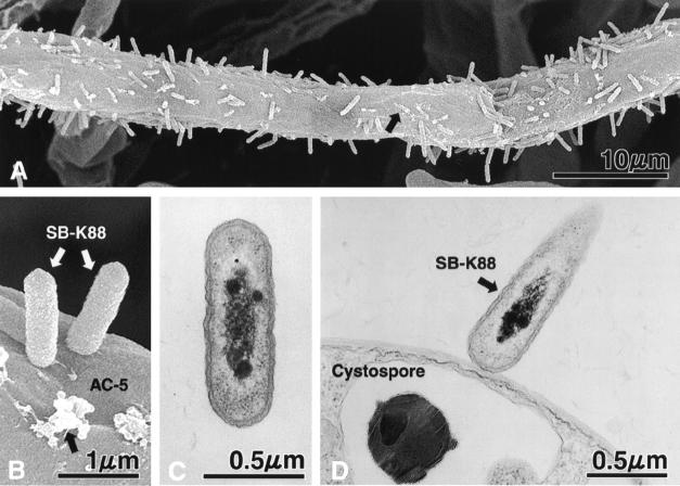
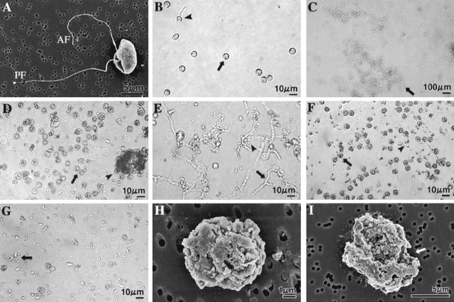
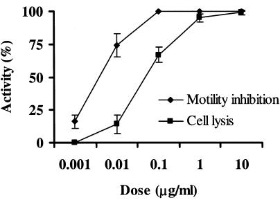
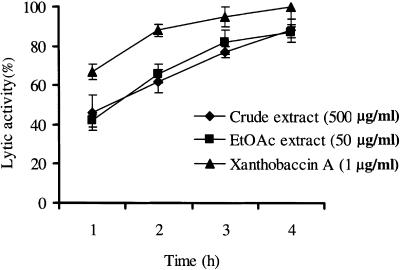
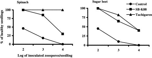
We previously demonstrated that xanthobaccin A from the rhizoplane bacterium Lysobacter sp. strain SB-K88 suppresses damping-off disease caused by Pythium sp. in sugar beet. In this study we focused on modes of Lysobacter sp. strain SB-K88 root colonization and antibiosis of the bacterium against Aphanomyces cochlioides, a pathogen of damping-off disease. Scanning electron microscopic analysis of 2-week-old sugar beet seedlings from seeds previously inoculated with SB-K88 revealed dense colonization on the root surfaces and a characteristic perpendicular pattern of Lysobacter colonization possibly generated via development of polar, brush-like fimbriae. In colonized regions a semitransparent film apparently enveloping the root and microcolonies were observed on the root surface. This Lysobacter strain also efficiently colonized the roots of several plants, including spinach, tomato, Arabidopsis thaliana, and Amaranthus gangeticus. Plants grown from both sugar beet and spinach seeds that were previously treated with Lysobacter sp. strain SB-K88 displayed significant resistance to the damping-off disease triggered by A. cochlioides. Interestingly, zoospores of A. cochlioides became immotile within 1 min after exposure to a SB-K88 cell suspension, a cell-free supernatant of SB-K88, or pure xanthobaccin A (MIC, 0.01 microg/ml). In all cases, lysis followed within 30 min in the presence of the inhibiting factor(s). Our data indicate that Lysobacter sp. strain SB-K88 has a direct inhibitory effect on A. cochlioides, suppressing damping-off disease. Furthermore, this inhibitory effect of Lysobacter sp. strain SB-K88 is likely due to a combination of antibiosis and characteristic biofilm formation at the rhizoplane of the host plant.


Figures

FIG. 1.
FIG. 1.
In vitro interactions between Lysobacter sp. strain SB-K88 and A. cochlioides AC-5 in a dual culture on PDA. (A) Inhibition of AC-5 mycelial growth in the presence of SB-K88 (arrow; 4 days). (B) Gliding motility of SB-K88 (arrow) on PDA (10 days) and changes in the hyphal density at the edge of the AC-5 colony. (C) Normal hyphal growth in a control. (D) Curly growth of AC-5 hyphae approaching an SB-K88 colony. The photographs in panels A and B were taken with a digital camera (CAMEDIA C-3040 zoom; Olympus Optical Co. Ltd.), and the micrographs in panels C and D were taken with the same digital camera connected to a light microscope (IX70-S1F2; Olympus).
FIG. 2.
FIG. 2.
Scanning electron micrographs of A. cochlioides mycelial samples interacting (5 days) with Lysobacter sp. strain SB-K88 and an untreated control. For the SEM study, small blocks (diameter, 6 mm) of affected mycelia (approaching a bacterial colony) in agar were transferred from a petri dish (inside diameter, 9 cm) containing corn meal agar to a 3-cm-inside-diameter petri dish on day 6 of cultivation and then fixed with 2% glutaraldehyde in phosphate buffer (8 mM, pH 7.2) for 3 h. Other preparation procedures for microscopy were similar to those described previously (14, 17, 18). (A) Normal growth in the absence of SB-K88 (control). (B) Bulbous structures (arrow) and curly growth in the presence of SB-K88. (C) Cytoplasmic extrusion from the hyphae (arrows). (D) Overlapping growth of mycelia (arrow).
FIG. 3.
FIG. 3.
Scanning electron micrographs showing seed coats of spinach (A and B) and sugar beet (C and D) at zero time (A and C) and 48 h (B and D) after inoculation of seeds with Lysobacter sp. strain SB-K88 cells (see Materials and Methods for details). The numbers of bacteria per 100 μm2 of seed coat determined by SEM were 69 ± 8 cells (zero time) and 158 ± 11 cells (48 h) for spinach and 22 ± 4 cells (zero time) and 53 ± 9 cells (48 h) for sugar beet. These values are averages ± standard errors of five replications.
FIG. 4.
FIG. 4.
TEM (A) and SEM (B to I) micrographs illustrating the morphology of Lysobacter sp. strain SB-K88 (A) and colonization of SB-K88 (B to I) on plant surfaces upon inoculation of seeds and seedlings grown in the gellan gum-based medium (B to D and G to I) or soil (F). (A) TEM micrograph of a sessile SB-K88 bacterial cell having large, brush-like fimbriae at one end. (B) Colonization on sugar beet root by perpendicular attachment. (C) Bacterial biofilm that developed under a semitransparent film of sugar beet root mucigel. (D) Typical perpendicular attachment of a bacterial cell to a sugar beet cotyledon. (E) High-density perpendicular attachment and colonization on the sugar beet leaf surface after immersion into an SB-K88 bacterial suspension (ca. 105 CFU/ml). (F) Colonization of sugar beet root. (G) Colonization of tomato root. (H) Colonization of A. thaliana leaf. (I) Colonization of A. thaliana root. Each experiment was repeated three times, and representative micrographs are shown (see Materials and Methods for details).
FIG. 5.
FIG. 5.
SEM (A and B) and TEM (C and D) micrographs showing perpendicular attachment of Lysobacter sp. strain SB-K88 to a hypha (A and B) and a cystospore (D) of A. cochlioides (for details see Materials and Methods) and a longitudinal section of SB-K88 (C). The black arrows in panels A and B indicate unidentified granular deposits on the surface of the A. cochlioides hyphae colonized by SB-K88. No such granular deposits were observed on the surfaces of untreated control hyphae (see Fig. 2A). Each experiment was repeated at least three times.
FIG. 6.
FIG. 6.
Light (B to G) and SEM (H and I) micrographs showing A. cochlioides zoospore-lytic activity of the freeze-dried culture supernatant, EtOAc- and water-soluble fractions of the culture supernatant, and pure xanthobaccin A. The micrographs in panels B to G were taken after 3 h of treatment by focusing on the bottom of a petri dish with a digital camera connected to the microscope (for details see Materials and Methods). Crude extracts or pure xanthobaccin A (dissolved in small quantities of DMSO) at the concentrations tested immediately caused inhibition of the motility of zoospores (see Table 1 and Materials and Method for details of the bioassay method). The halted zoospores rapidly settled to the bottom of the dish and then started to burst or lyse. The final concentration of DMSO in the aqueous zoospore suspension was maintained at less than 1% in all treatments. DMSO alone (final concentration, 1%) was used as the negative control and caused no lysis of zoospores. Each experiment was replicated at least five times, and representative micrographs are shown. (A) SEM micrograph of a biflagellate A. cochlioides zoospore (untreated control). AF, anterior flagellum; PF, posterior flagellum. (B) No lysis in the control dish (1% DMSO). A small portion (10 to 15%) of the motile zoospores in the control dish were stopped and changed into round cystospores (arrow) after 3 h and then settled to the bottom of the dish; 5 to 8% of these cystospores germinated (arrowhead) and formed germ tubes. No motile zoospores were observed in aqueous medium because the photograph was taken by focusing on bottom of the dish. (C) Complete lysis of all halted zoospores by freeze-dried culture supernatant (500 μg/ml). The arrow indicates lysed material. (D) All spores were granulated or lysed (arrow) by the EtOAc-soluble fraction (100 μg/ml). Some lysed material aggregated (arrowhead). (E) Water-soluble fraction (500 μg/ml) initially induced germination of cystospores (arrow and arrowhead) within 1 h, and then (3 h) all spores and germ tubes were partially lysed (arrow and arrowhead). (F) Xanthobaccin A (1 μg/ml) caused granulation (arrowhead) and lysis (arrowhead) of all spores. (G) Complete lysis of zoospores (arrow) by xanthobaccin A (1 μg/ml). (H and I) Scanning electron micrographs of granulated, cracked, and lysed (arrow in I) A. cochlioides zoospores exposed to 1 ppm xanthobaccin A for 30 min.
FIG. 7.
FIG. 7.
Inhibition of motility and lysis of A. cochlioides zoospores treated with various doses of xanthobaccin A. Xanthobaccin A was first dissolved in a small quantity of DMSO and then serially diluted with distilled water. Appropriate amounts of a sample suspension were added to the aqueous zoospore suspension. The final DMSO concentration was always less than 1% in the zoospore suspension. The inhibition of motility and lysis of zoospores were observed microscopically (magnification, ×20) (for details see Table 1) 3 h after the treatment with xanthobaccin A, and the percentages of activity were calculated as described previously (16). DMSO alone (final concentration in the zoospore suspension, 1%) was used as the control and did not have any effect on the motility and lysis of the zoospores. The data are the averages ± standard errors of at least three replications for each dose of xanthobaccin A.
FIG. 8.
FIG. 8.
Time course of lytic activity of the crude freeze-dried culture supernatant, the EtOAc-soluble fraction, and xanthobaccin A against A. cochlioides zoospores. Xanthobaccin A or extracts of the culture supernatant were first dissolved in a small quantity of DMSO and then serially diluted with distilled water. Appropriate amounts of a sample suspension were added to the aqueous zoospore suspension. The final DMSO concentration was always less than 1% in the zoospore suspension. The time course of lysis of zoospores was observed microscopically (magnification, ×20) (for details see Table 1) for up to 4 h after the treatment, and the percentages of activity were calculated as described previously (16). DMSO alone (final concentration in the zoospore suspension, 1%) was used as the control and did not cause any lysis of the zoospores. The data are the averages ± standard errors of at least three replications for each dose of xanthobaccin A.
FIG. 9.
FIG. 9.
Damping-off disease suppression by Lysobacter sp. strain SB-K88 in spinach and sugar beet. Five surface-sterilized seeds coated with nutrient-free SB-K88 cells (ca. 108 CFU/seed) or fungicide (Tachigaren; 7.5 g/kg seeds) were sown in 36-cell plastic packs containing sterilized soil. On day 12 of cultivation, each seedling was inoculated with an appropriate number of zoospores (for details see Materials and Methods). The percentage of healthy seedlings was recorded 2 weeks after zoospore inoculation. Seedlings that did not have black and shrunken or dark, slender hypocotyls or roots were considered healthy seedlings. Seedlings obtained from surface-sterilized seeds (no treatment with bacteria or fungicide) were considered controls. The values are averages of three replications.

References

    1. Bais, H. P., R. Fall, and J. M. Vivanco. 2004. Biocontrol of Bacillus subtilis against infection of Arabidopsis root by Pseudomonas syringae is facilitated by biofilm formation and surfactin production. Plant Physiol. 134:307-319. - PMC - PubMed
    1. Bonner, D. P., J. O'Sullivan, S. K. Tanaka, J. M. Clark, and R. R. Whitney. 1988. Lysobactin, a novel antibacterial agent produced by Lysobacter sp. II. Biological properties. J. Antibiot. 41:1745-1751. - PubMed
    1. Chin-A-Woeng, T. F. C., W. D. Priester, A. J. V. D. Bij, and B. J. J. Lugtenburg. 1997. Description of the colonization of a gnotobiotic tomato rhizosphere by Pseudomonas fluorescens biocontrol strain WCS365, using scanning electron microscopy. Mol. Plant-Microbe Interact. 10:79-86.
    1. Christensen, P., and F. D. Cook. 1978. Lysobacter, a new genus of non-fruiting, gliding bacteria with high base ratio. Int. J. Syst. Bacteriol. 28:367-393.
    1. Cook, R. J., L. S. Thomashow, D. M. Weller, D. Fujimoto, M. Mazzola, G. Bangera, and D.-S. Kim. 1995. Molecular mechanisms of defense by rhizobacteria against root disease. Proc. Natl. Acad. Sci. USA 92:4197-4201. - PMC - PubMed

Publication types

MeSH terms

LinkOut - more resources