. 2005 Jul;71(7):4057-68.
doi: 10.1128/AEM.71.7.4057-4068.2005.

Genomic organization and molecular analysis of virulent bacteriophage 2972 infecting an exopolysaccharide-producing Streptococcus thermophilus strain

Affiliations

Genomic organization and molecular analysis of virulent bacteriophage 2972 infecting an exopolysaccharide-producing Streptococcus thermophilus strain

Céline Lévesque et al. Appl Environ Microbiol. 2005 Jul.

Abstract

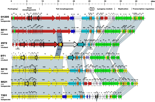
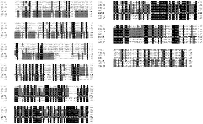
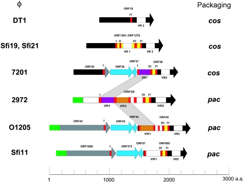
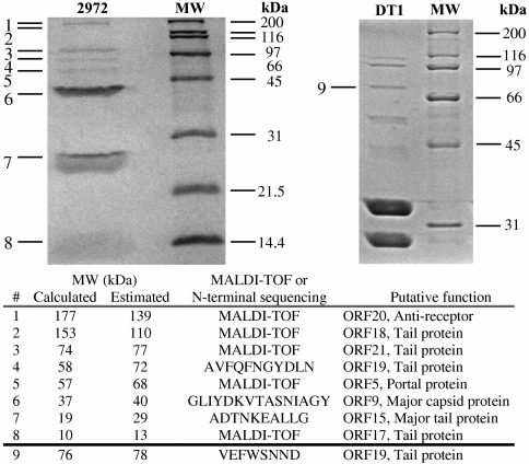
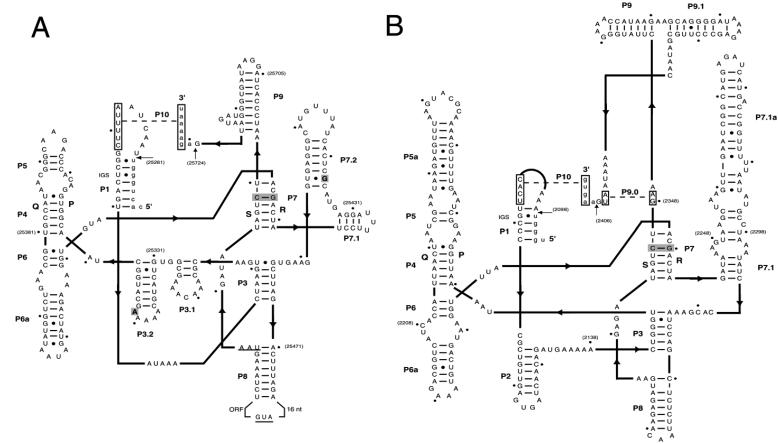
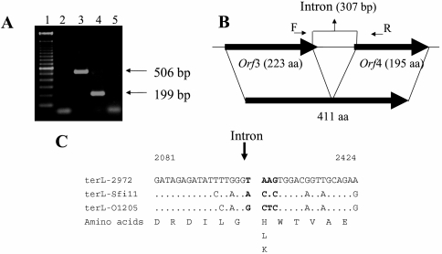
The Streptococcus thermophilus virulent pac-type phage 2972 was isolated from a yogurt made in France in 1999. It is a representative of several phages that have emerged with the industrial use of the exopolysaccharide-producing S. thermophilus strain RD534. The genome of phage 2972 has 34,704 bp with an overall G+C content of 40.15%, making it the shortest S. thermophilus phage genome analyzed so far. Forty-four open reading frames (ORFs) encoding putative proteins of 40 or more amino acids were identified, and bioinformatic analyses led to the assignment of putative functions to 23 ORFs. Comparative genomic analysis of phage 2972 with the six other sequenced S. thermophilus phage genomes confirmed that the replication module is conserved and that cos- and pac-type phages have distinct structural and packaging genes. Two group I introns were identified in the genome of 2972. They interrupted the genes coding for the putative endolysin and the terminase large subunit. Phage mRNA splicing was demonstrated for both introns, and the secondary structures were predicted. Eight structural proteins were also identified by N-terminal sequencing and/or matrix-assisted laser desorption ionization-time-of-flight mass spectrometry. Detailed analysis of the putative minor tail proteins ORF19 and ORF21 as well as the putative receptor-binding protein ORF20 showed the following interesting features: (i) ORF19 is a hybrid protein, because it displays significant identity with both pac- and cos-type phages; (ii) ORF20 is unique; and (iii) a protein similar to ORF21 of 2972 was also found in the structure of the cos-type phage DT1, indicating that this structural protein is present in both S. thermophilus phage groups. The implications of these findings for phage classification are discussed.


Figures

FIG. 1.
FIG. 1.
Dendrogram of EcoRV restriction profiles of 12 virulent phages infecting S. thermophilus RD534. The phage names and the years and countries of isolation are given. Each phage was isolated from a different dairy factory.
FIG. 2.
FIG. 2.
Electron micrograph of S. thermophilus phage 2972 virions negatively stained with 2% phosphotungstic acid (pH 7.2). Bar, 100 nm.
FIG. 3.
FIG. 3.
Alignment of the genetic maps of all completely sequenced S. thermophilus phage genomes. The modular regions of the genomes coding for distinct functions are indicated above the maps. Deduced proteins sharing more than 50% amino acid identity are represented using the same color and are linked using grey shading whenever possible. ORFs with unique sequences are displayed in white. Genes coding for proteins identified by N-terminal sequencing or MALDI-TOF are identified by thick lines.
FIG. 4.
FIG. 4.
Alignment of ORF19 of phage 2972 with similar proteins found in S. thermophilus phages 7201, Sfi21, Sfi19, DT1, Sfi11, and O1205. Amino acids conserved in six or seven aligned sequences are identified by black shading. Amino acids that are conserved in five or fewer sequences are identified by gray shading.
FIG. 5.
FIG. 5.
Schematic illustration representing the alignment of the proteins possibly involved in host recognition of seven S. thermophilus phages. The same color indicates more than 80% similarity. Collagen-like repeats are shown in red. VR1, VR2, and VR3 indicate variable regions 1, 2, and 3, respectively. Regions with unique sequences are shown in white.
FIG. 6.
FIG. 6.
Protein profiles of phages 2972 (pac type) and DT1 (cos type) as determined on SDS-PAGE gels stained with Coomassie blue. MW, molecular weight markers.
FIG. 7.
FIG. 7.
Secondary-structure predictions of the two introns in the genome of phage 2972. The secondary-structure representation was made using a two-dimensional structural diagram (15, 18, 46). Arrows indicate the 5′ and 3′ splicing sites. Lower- and uppercase letters denote the exon and intron sequences, respectively. Boxed sequences indicate the regions that anneal to form P9.0 and P10. The shaded nucleotides in P7 represent the putative guanosine-binding site. Bold lines show connections between intron structure domains, with pointers indicating the 5′-to-3′ direction. IGS, internal guide sequence. Numbers in parentheses represent the nucleotide position on the phage 2972 genome. (A) Intron in the gene coding for the endolysin. The start and stop codons of ORF28 are underlined, and the two nucleotides that differ from the S3b intron are boldfaced. (B) Intron in the gene coding for the large subunit of the terminase.
FIG. 8.
FIG. 8.
Characterization of the intron between the genes coding for the terminase large subunit of phage 2972. (A) In vivo splicing of 2972 intron RNA (terminase large-subunit gene). Lane 1, 100-bp marker (Invitrogen); lane 2, negative control without DNA, cDNA, or RNA; lane 3, PCR product obtained with 2972 DNA as a template; lane 4, PCR product obtained with RNA isolated from 2972-infected S. thermophilus cells (cDNA); lane 5, PCR product (no reverse transcription) obtained with RNA isolated from 2972-infected cells. (B) Phage 2972 genomic region containing the intron and the two ORFs coding for the terminase large subunit. Large, thick arrows indicate open reading frames; the numbers of amino acids (aa) in the deduced proteins are given. Splicing of the 307-bp intron resulted in a 411-aa protein. Small arrows represent the primers used for PCR and sequencing. (C) Nucleotide sequence alignment of the regions flanking the splicing site with the corresponding intron-free regions in two other pac-type phages (Sfi11 [orf411] and O1205 [orf26]). Differences relative to the phage 2972 sequence are indicated. Vertical arrow indicates the intron insertion site. The nucleotide positions are based on the 2972 genomic sequence (GenBank accession no. AY699705).

References

    1. Ackermann, H.-W. 1999. Tailed bacteriophages: the order Caudovirales. Adv. Virus Res. 51:135-201. - PMC - PubMed
    1. Altschul, S. F., T. L. Madden, A. A. Schaffer, J. Zhang, Z. Zhang, W. Miller, and D. J. Lipman. 1997. Gapped BLAST and PSI-BLAST: a new generation of protein database search programs. Nucleic Acids Res. 25:3389-3402. - PMC - PubMed
    1. Bechhofer, D. H., K. K. Hue, and D. A. Shub. 1994. An intron in the thymidylate synthase gene of Bacillus bacteriophage β22: evidence for independent evolution of a gene, its group I intron, and the intron open reading frame. Proc. Natl. Acad. Sci. USA 91:11669-11673. - PMC - PubMed
    1. Beck, K., and B. Brodsky. 1998. Supercoiled protein motifs: the collagen triple-helix and the α-helical coiled coil. J. Struct. Biol. 122:17-29. - PubMed
    1. Beres, S. B., G. L. Sylva, K. D. Barbian, B. Lei, J. S. Hoff, N. D. Mammarelle, M.-Y. Liu, J. C. Smoot, S. F. Porcella, L. D. Parkins, D. S. Campbell, T. M. Smith, J. K. McCormick, D. Y. M. Leung, P. M. Schlievert, and J. M. Musser. 2002. Genome sequence of a serotype M3 strain of a group A Streptococcus: phage-encoded toxins, the high-virulence phenotype, and clone emergence. Proc. Natl. Acad. Sci. USA 99:10078-10083. - PMC - PubMed

Publication types

Associated data